Block-copolymer-stabilized iodinated emulsions for use as CT contrast agents Anke de Vries a , Erica Custers b , Johan Lub b , Sandra van den Bosch b , Klaas Nicolay b , Holger Grüll a, b, * a Biomedical NMR, Department of Biomedical Engineering, Eindhoven University of Technology, Den Dolech 2, 5600 MB Eindhoven, The Netherlands b Department of Biomolecular Engineering, Philips Research Eindhoven, HTC 11, 5656 AE Eindhoven, The Netherlands article info Article history: Received 2 February 2010 Accepted 27 April 2010 Available online 11 June 2010 Keywords: Computed tomography (CT) Contrast agent Emulsion Nanoparticles Iodine X-ray imaging abstract The objective of this study was to develop radiopaque iodinated emulsions for use as CT blood pool contrast agents. Three hydrophobic iodinated oils were synthesized based on the 2,3,5-triiodobenzoate moiety and formulated into emulsions using either phospholipids or amphiphilic polymers, i.e. Pluronic F68 and poly(butadiene)-b-poly(ethylene glycol) (PBD-PEO), as emulsifiers. The size, stability and cell viability was investigated for all stabilized emulsions. Three emulsions stabilized with either lipids or PBD-PEO were subsequently tested in vivo as a CT blood pool contrast agent in mice. While the lipid- stabilized emulsions turned out unstable in vivo, polymer-stabilized emulsions performed well in vivo. In blood, a contrast enhancement of 220 Hounsfield Units (HU) was measured directly after intravenous administration of 520 mg I/kg. The blood circulation half-life of a PBD-PEO stabilized emulsion was approximately 3 h and no noticeable in vivo toxicity was observed. These results show the potential of above emulsions for use as blood pool agents in contrast enhanced CT imaging. Ó 2010 Elsevier Ltd. All rights reserved. 1. Introduction X-ray and computed tomography (CT) are the most frequently used diagnostic imaging technologies in the clinic. Recent techno- logical advances, such as fast digital X-ray detectors or spiral and multi-slice CT [1,2], in combination with the approval of improved iodinated CT contrast agents, opened a plethora of new applications in radiology. With X-ray and CT moving forward into the inter- ventional care, such as stent placement, balloon dilatation, vascular surgery, and electrophysiology procedures, there is a clear need for CT contrast agents that allow sharp blood vessel delineation and have a long circulation time to avoid multiple CA injections [3,4]. Current contrast agents used in X-ray and CT applications are usually iodinated molecules with a low molecular weight (<2000 Da) resulting in a rapid renal excretion and a high free volume of distribution as they rapidly equilibrate between the blood compartment and the extracellular, extravascular compart- ment [5,6]. One strategy to improve the current generation of CT contrast agents is to design a blood pool CT contrast agents having sizes larger than ca. 5.5 nm to prohibit rapid renal excretion and extravasation [7]. Blood clearance of these agents occurs via uptake in the reticulo-endothelial system (RES) followed by metabolism and/or excretion via the hepatobiliary pathway. Long circulation times can be achieved by designing a stealth coating on the agents’ surface to avoid rapid opsonization followed by macrophage uptake. Moreover, it is desired that the CT contrast agent has a high iodine payload to avoid injection of a large volume. The latter is of special interest for preclinical studies using CT contrast agents as the maximum volume that can be injected intravenously into mice is generally restricted to w200 mL. Whereas most CT contrast agents are based on the high-Z element iodine, also other high-Z elements have been explored for their radiopaque properties in preclinical studies. For example, nanoparticles based on bismuth sulphide (10e50 nm) or gold (38 nm) with blood half-lives of 140 15 min and 14.6 3.3 h respectively were investigated in mice [8,9]. So far, a detailed study on the toxicity of bismuth-based nanoparticles is lacking, however, several studies report on the potential toxic effect of gold nano- particles [10e12], which hampers their translation into the clinic. Most investigations on new CT contrast agents focus on iodine due to similarities with the already clinical approved agents. Margel and coworkers designed solid nanoparticles of 30.6 5 nm for X-ray imaging based on polymers of 2-methacryloyloxyethy(2,3,5-trii- dobenzoate) [13e15], which is a similar approach taken by Hyafil et al. [16]. Besides highly radio-opaque solid nanoparticles, also iodinated polymeric micelles of MPEG-iodolysine block-copoly- mers [17,18] and emulsions of iodinated triglycerides were inves- tigated [19]. The latter is commercially available for preclinical use * Corresponding author at: Eindhoven University of Technology, Dept. of Biomedical Engineering, Biomedical NMR, Den Dolech 2 (N-laag 2.62), 5600 MB Eindhoven, The Netherlands. Tel.: þ31 (0) 612932309; fax: þ31 (0) 402744906. E-mail address: [email protected](H. Grüll). Contents lists available at ScienceDirect Biomaterials journal homepage: www.elsevier.com/locate/biomaterials 0142-9612/$ e see front matter Ó 2010 Elsevier Ltd. All rights reserved. doi:10.1016/j.biomaterials.2010.04.056 Biomaterials 31 (2010) 6537e6544
Transcript
lable at ScienceDirect
Biomaterials 31 (2010) 6537e6544
Contents lists avai
Biomaterials
journal homepage: www.elsevier .com/locate/biomater ia ls
Block-copolymer-stabilized iodinated emulsions for use as CT contrast agents
Anke de Vries a, Erica Custers b, Johan Lub b, Sandra van den Bosch b, Klaas Nicolay b, Holger Grüll a,b,*aBiomedical NMR, Department of Biomedical Engineering, Eindhoven University of Technology, Den Dolech 2, 5600 MB Eindhoven, The NetherlandsbDepartment of Biomolecular Engineering, Philips Research Eindhoven, HTC 11, 5656 AE Eindhoven, The Netherlands
a r t i c l e i n f o
Article history:Received 2 February 2010Accepted 27 April 2010Available online 11 June 2010
0142-9612/$ e see front matter � 2010 Elsevier Ltd.doi:10.1016/j.biomaterials.2010.04.056
a b s t r a c t
The objective of this study was to develop radiopaque iodinated emulsions for use as CT blood poolcontrast agents. Three hydrophobic iodinated oils were synthesized based on the 2,3,5-triiodobenzoatemoiety and formulated into emulsions using either phospholipids or amphiphilic polymers, i.e. PluronicF68 and poly(butadiene)-b-poly(ethylene glycol) (PBD-PEO), as emulsifiers. The size, stability and cellviability was investigated for all stabilized emulsions. Three emulsions stabilized with either lipids orPBD-PEO were subsequently tested in vivo as a CT blood pool contrast agent in mice. While the lipid-stabilized emulsions turned out unstable in vivo, polymer-stabilized emulsions performed well in vivo. Inblood, a contrast enhancement of 220 Hounsfield Units (HU) was measured directly after intravenousadministration of 520 mg I/kg. The blood circulation half-life of a PBD-PEO stabilized emulsion wasapproximately 3 h and no noticeable in vivo toxicity was observed. These results show the potential ofabove emulsions for use as blood pool agents in contrast enhanced CT imaging.
� 2010 Elsevier Ltd. All rights reserved.
1. Introduction
X-ray and computed tomography (CT) are the most frequentlyused diagnostic imaging technologies in the clinic. Recent techno-logical advances, such as fast digital X-ray detectors or spiral andmulti-slice CT [1,2], in combination with the approval of improvediodinated CTcontrast agents, opened a plethora of newapplicationsin radiology. With X-ray and CT moving forward into the inter-ventional care, such as stent placement, balloon dilatation, vascularsurgery, and electrophysiology procedures, there is a clear need forCT contrast agents that allow sharp blood vessel delineation andhave a long circulation time to avoid multiple CA injections [3,4].Current contrast agents used in X-ray and CT applications areusually iodinated molecules with a low molecular weight(<2000 Da) resulting in a rapid renal excretion and a high freevolume of distribution as they rapidly equilibrate between theblood compartment and the extracellular, extravascular compart-ment [5,6]. One strategy to improve the current generation of CTcontrast agents is to design a blood pool CT contrast agents havingsizes larger than ca. 5.5 nm to prohibit rapid renal excretion andextravasation [7]. Blood clearance of these agents occurs via uptake
ty of Technology, Dept. ofh 2 (N-laag 2.62), 5600 MB; fax: þ31 (0) 402744906.
All rights reserved.
in the reticulo-endothelial system (RES) followed by metabolismand/or excretion via the hepatobiliary pathway. Long circulationtimes can be achieved by designing a stealth coating on the agents’surface to avoid rapid opsonization followed by macrophageuptake. Moreover, it is desired that the CT contrast agent has a highiodine payload to avoid injection of a large volume. The latter is ofspecial interest for preclinical studies using CT contrast agents asthe maximum volume that can be injected intravenously into miceis generally restricted to w200 mL.
Whereas most CT contrast agents are based on the high-Zelement iodine, also other high-Z elements have been explored fortheir radiopaque properties in preclinical studies. For example,nanoparticles based on bismuth sulphide (10e50 nm) or gold(38 nm) with blood half-lives of 140 � 15 min and 14.6 � 3.3 hrespectively were investigated in mice [8,9]. So far, a detailed studyon the toxicity of bismuth-based nanoparticles is lacking, however,several studies report on the potential toxic effect of gold nano-particles [10e12], which hampers their translation into the clinic.Most investigations on new CT contrast agents focus on iodine dueto similarities with the already clinical approved agents. Margel andcoworkers designed solid nanoparticles of 30.6 � 5 nm for X-rayimaging based on polymers of 2-methacryloyloxyethy(2,3,5-trii-dobenzoate) [13e15], which is a similar approach taken by Hyafilet al. [16]. Besides highly radio-opaque solid nanoparticles, alsoiodinated polymeric micelles of MPEG-iodolysine block-copoly-mers [17,18] and emulsions of iodinated triglycerides were inves-tigated [19]. The latter is commercially available for preclinical use
A. de Vries et al. / Biomaterials 31 (2010) 6537e65446538
(Fenestra VC (ART, Saint-Laurent, Canada)), however, the low iodineconcentration of 50 mg I/mL hampers its use in small animals dueto restrictions on the injectable volume.
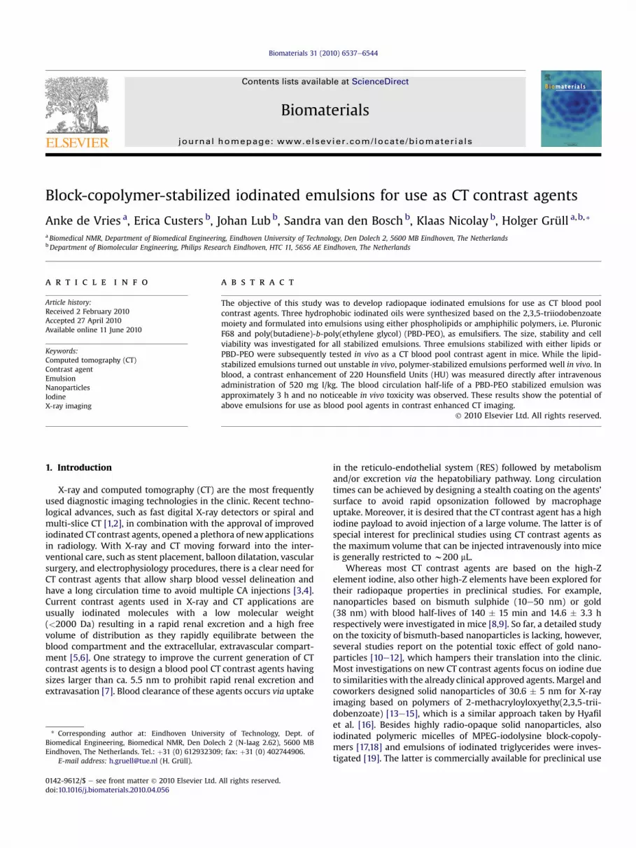
In our work, we explored highly iodinated nano-emulsions asa possible class of blood pool CT contrast agent. The radiopaquecore of the emulsions consists of a hydrophobic iodinated oil, e.g.octan-2-yl 2,3,5-triiodobenzoate (oil 1), 3,7-dimethyloctyl 2,3,5-triiodobenzoate (oil 2), or 2,3,5-triiodobenzyl 2-methylheptanoate(oil 3). These oils were emulsified using three types of surfactants,namely 1,2-distearoyl-sn-glycero-3-phosphocholine (DSPC; a phos-pholipid), poly(ethylene oxide)-b-poly(propylene oxide)-b-poly(ethylene oxide) (PEO-PPO-PEO; Pluronic F68) or poly(butadiene)-b-poly(ethylene oxide) (PBD-PEO) (Fig. 1). The obtained CT contrastagents were characterized in great detail using dynamic lightscattering, Coulter Counter, and Cryo-TEM. MTT toxicity assayswere performed to test for acute cell toxicity. Finally, the bestcandidate emulsions (DSPC, oil 1; PBD-PEO, oil 1; PBD-PEO, oil 2)were taken into in vivo tests to study blood half-life and contrastgeneration in CT imaging of mice.
2. Experimental
2.1. Materials
All solventswereobtained fromMerck.2-Methylheptanoicacidwasobtained fromAcros. 1,2-Distearoyl-sn-glycero-3-phosphocholine (DSPC), 1,2-distearoyl-sn-Glycero-3-phospho-ethanolamine-N-[methoxy(polyethylene glycol)-2000] (PEG2000-DSPE)were purchased from Lipoid and cholesterol (Chol) was purchased from AvantiPolar Lipids. Pluronic F-68 (PEO-PPO-PEO:Mw,PEO¼ 3360 g/mol;Mw,PPO¼ 1680 g/mol;Mw,PEO ¼ 3360 g/mol) [20] was purchased from SigmaeAldrich. Poly(butadiene(1,2-addition)-b-ethylene oxide) (P-6088;Mw,PEO¼ 2033 g/mol;Mw,PBD¼ 1305 g/mol) wasobtained from Polymer Source. All other commercial chemicals were purchased fromSigmaeAldrich in the highest available quality.
2.2. Synthesis
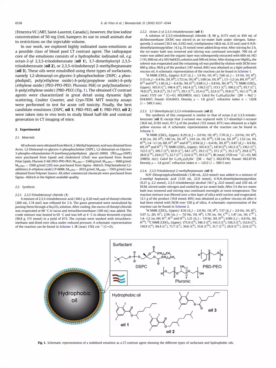
2.2.1. 2,3,5-Triiodobenzoyl chloride (1)Amixture of 2,3,5-triiodobenzoic acid (100.1 g, 0.20 mol) and of thionyl chloride
(200 mL, 1.74 mol) was refluxed for 3 h. The gases generated were neutralized bypassing them through a Na2CO3 solution. After cooling, the excess of thionyl chloridewas evaporated at 60 �C in vacuo and tetrachloromethane (100 mL) was added. Thecrude mixture was heated to 65 �C and was left at 4 �C to obtain brownish crystals(90.8 g, 175 mmol) in a yield of 87%. The crystals were washed with tetrachloro-methane and dried over silica under reduced pressure. A schematic representationof the reaction can be found in Scheme 1. IR (neat) 1762 cm�1 (C]O).
Fig. 1. Schematic representation of a stabilized emulsion as a CT contrast
2.2.2. Octan-2-yl 2,3,5-triiodobenzoate (oil 1)A solution of 2,3,5-triiodobenzoyl chloride (1, 90 g, 0.175 mol) in 450 mL of
dichloromethane (DCM) was stirred in an ice-water bath under nitrogen. Subse-quently 2-octanol (30.5 mL, 0.192 mol), triethylamine (48.8 mL, 0.35 mol) and 4-N,N-dimethylaminopyridine (4.3 g, 35mmol) were added dropwise. After stirring for 2 h,the ice-water bath was removed and stirring was continued overnight. 700 mL ofwater was added and the organic layer was subsequently extracted with 600 mL HCl(1N), 600mLof a 10%NaHCO3 solution and500mLbrine.AfterdryingoverMgSO4, thesolvent was evaporated and the remaining oil was purified by elutionwith DCM over660 g silica. 90.0 g of the product (147 mmol, 84%) was obtained as a light yellowishviscous oil. A schematic representation of the reaction can be found in Scheme 1.
2.2.3. 3,7-Dimethyloctyl 2,3,5-triiodobenzoate (oil 2)The synthesis of this compound is similar to that of octan-2-yl 2,3,5-triiodo-
benzoate (oil 1) except that 2-octanol was replaced with 3,7-dimethyl-1-octanol(36.8 mL, 0.192 mol). 97.7 g of the product (152 mmol, 87%) was obtained as a lightyellow viscous oil. A schematic representation of the reaction can be found inScheme 1.
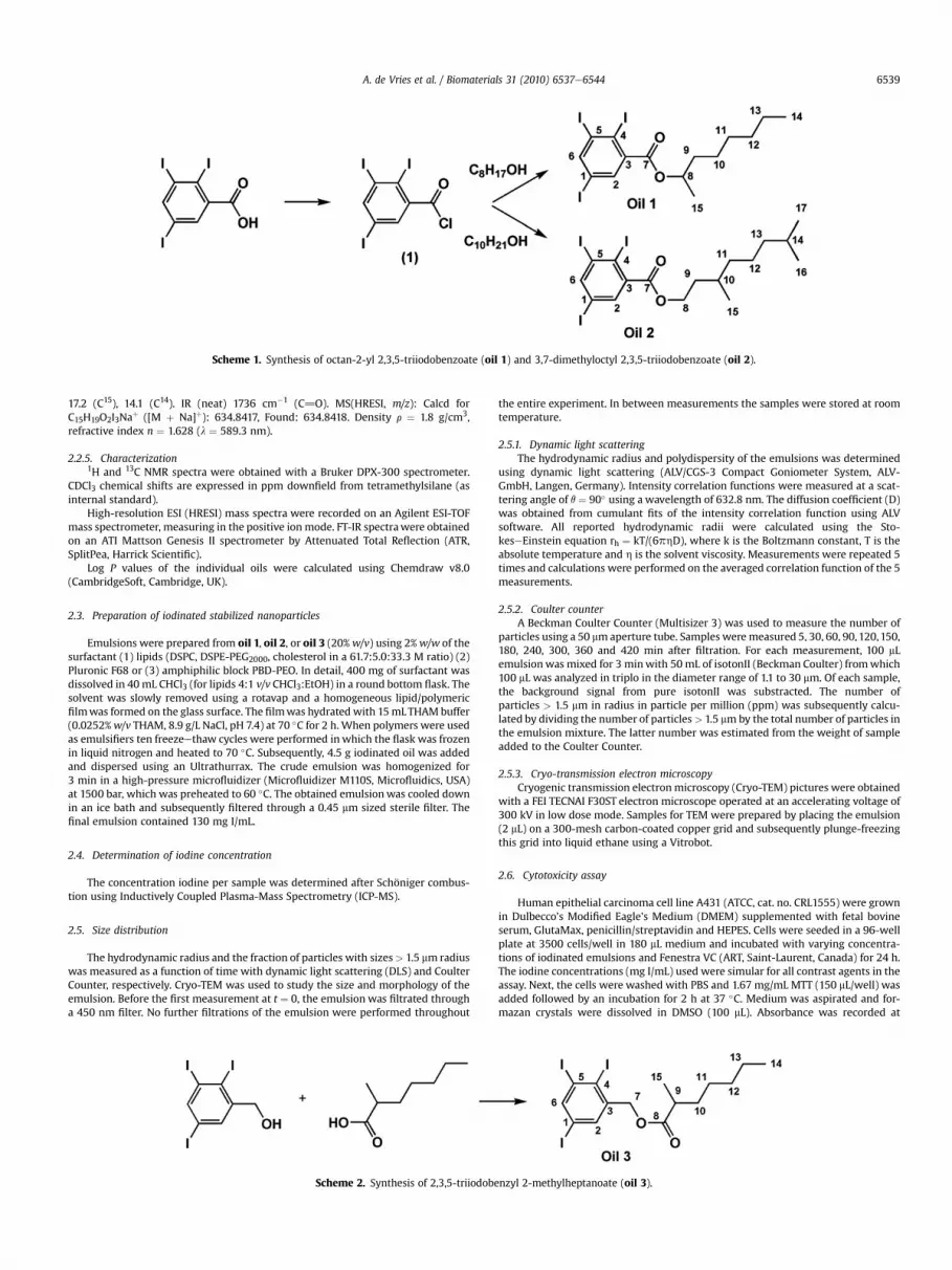
2.2.4. 2,3,5-Triiodobenzyl 2-methylheptanoate (oil 3)N,N0-Diisopropylcarbodiimide (3.40 mL, 22.0 mmol) was added to a mixture of
2-methyl heptanoic acid (3.50 mL, 22.0 mmol), 4-N,N-dimethylaminopyridine(0.27 g, 2.2 mmol), 2,3,5-triiodobenzyl alcohol (10.7 g, 22.0 mmol) and 250 mL ofDCM, stirred under nitrogen and cooled by an ice-water bath. After 2 h the ice-waterbath was removed and stirring was continued overnight at room temperature. Thereaction mixture was filtered over a thin layer of silica with suction and evaporated.12.1 g of the product (19.8 mmol, 89%) was obtained as a yellow viscous oil after ithad been eluted with DCM over 330 g of silica. A schematic representation of thereaction can be found in Scheme 2.
agent showing the different types of surfactant and hydrophobic oils.
Scheme 1. Synthesis of octan-2-yl 2,3,5-triiodobenzoate (oil 1) and 3,7-dimethyloctyl 2,3,5-triiodobenzoate (oil 2).
A. de Vries et al. / Biomaterials 31 (2010) 6537e6544 6539
17.2 (C15), 14.1 (C14). IR (neat) 1736 cm�1 (C]O). MS(HRESI, m/z): Calcd forC15H19O2I3Naþ ([M þ Na]þ): 634.8417, Found: 634.8418. Density r ¼ 1.8 g/cm3,refractive index n ¼ 1.628 (l ¼ 589.3 nm).
2.2.5. Characterization1H and 13C NMR spectra were obtained with a Bruker DPX-300 spectrometer.
CDCl3 chemical shifts are expressed in ppm downfield from tetramethylsilane (asinternal standard).
High-resolution ESI (HRESI) mass spectra were recorded on an Agilent ESI-TOFmass spectrometer, measuring in the positive ion mode. FT-IR spectrawere obtainedon an ATI Mattson Genesis II spectrometer by Attenuated Total Reflection (ATR,SplitPea, Harrick Scientific).
Log P values of the individual oils were calculated using Chemdraw v8.0(CambridgeSoft, Cambridge, UK).
2.3. Preparation of iodinated stabilized nanoparticles
Emulsions were prepared from oil 1, oil 2, or oil 3 (20%w/v) using 2%w/w of thesurfactant (1) lipids (DSPC, DSPE-PEG2000, cholesterol in a 61.7:5.0:33.3 M ratio) (2)Pluronic F68 or (3) amphiphilic block PBD-PEO. In detail, 400 mg of surfactant wasdissolved in 40 mL CHCl3 (for lipids 4:1 v/v CHCl3:EtOH) in a round bottom flask. Thesolvent was slowly removed using a rotavap and a homogeneous lipid/polymericfilmwas formed on the glass surface. The filmwas hydrated with 15mLTHAMbuffer(0.0252%w/v THAM, 8.9 g/L NaCl, pH 7.4) at 70 �C for 2 h. When polymers were usedas emulsifiers ten freezeethaw cycles were performed in which the flask was frozenin liquid nitrogen and heated to 70 �C. Subsequently, 4.5 g iodinated oil was addedand dispersed using an Ultrathurrax. The crude emulsion was homogenized for3 min in a high-pressure microfluidizer (Microfluidizer M110S, Microfluidics, USA)at 1500 bar, which was preheated to 60 �C. The obtained emulsionwas cooled downin an ice bath and subsequently filtered through a 0.45 mm sized sterile filter. Thefinal emulsion contained 130 mg I/mL.
2.4. Determination of iodine concentration
The concentration iodine per sample was determined after Schöniger combus-tion using Inductively Coupled Plasma-Mass Spectrometry (ICP-MS).
2.5. Size distribution
The hydrodynamic radius and the fraction of particles with sizes> 1.5 mm radiuswas measured as a function of time with dynamic light scattering (DLS) and CoulterCounter, respectively. Cryo-TEM was used to study the size and morphology of theemulsion. Before the first measurement at t ¼ 0, the emulsion was filtrated througha 450 nm filter. No further filtrations of the emulsion were performed throughout
Scheme 2. Synthesis of 2,3,5-triiodobe
the entire experiment. In between measurements the samples were stored at roomtemperature.
2.5.1. Dynamic light scatteringThe hydrodynamic radius and polydispersity of the emulsions was determined
using dynamic light scattering (ALV/CGS-3 Compact Goniometer System, ALV-GmbH, Langen, Germany). Intensity correlation functions were measured at a scat-tering angle of q ¼ 90� using a wavelength of 632.8 nm. The diffusion coefficient (D)was obtained from cumulant fits of the intensity correlation function using ALVsoftware. All reported hydrodynamic radii were calculated using the Sto-keseEinstein equation rh ¼ kT/(6phD), where k is the Boltzmann constant, T is theabsolute temperature and h is the solvent viscosity. Measurements were repeated 5times and calculations were performed on the averaged correlation function of the 5measurements.
2.5.2. Coulter counterA Beckman Coulter Counter (Multisizer 3) was used to measure the number of
particles using a 50 mmaperture tube. Samples weremeasured 5, 30, 60, 90,120,150,180, 240, 300, 360 and 420 min after filtration. For each measurement, 100 mLemulsionwas mixed for 3 minwith 50 mL of isotonII (Beckman Coulter) fromwhich100 mL was analyzed in triplo in the diameter range of 1.1 to 30 mm. Of each sample,the background signal from pure isotonII was substracted. The number ofparticles > 1.5 mm in radius in particle per million (ppm) was subsequently calcu-lated by dividing the number of particles> 1.5 mmby the total number of particles inthe emulsion mixture. The latter number was estimated from the weight of sampleadded to the Coulter Counter.
2.5.3. Cryo-transmission electron microscopyCryogenic transmission electronmicroscopy (Cryo-TEM) pictures were obtained
with a FEI TECNAI F30ST electron microscope operated at an accelerating voltage of300 kV in low dose mode. Samples for TEM were prepared by placing the emulsion(2 mL) on a 300-mesh carbon-coated copper grid and subsequently plunge-freezingthis grid into liquid ethane using a Vitrobot.
2.6. Cytotoxicity assay
Human epithelial carcinoma cell line A431 (ATCC, cat. no. CRL1555) were grownin Dulbecco’s Modified Eagle’s Medium (DMEM) supplemented with fetal bovineserum, GlutaMax, penicillin/streptavidin and HEPES. Cells were seeded in a 96-wellplate at 3500 cells/well in 180 mL medium and incubated with varying concentra-tions of iodinated emulsions and Fenestra VC (ART, Saint-Laurent, Canada) for 24 h.The iodine concentrations (mg I/mL) used were simular for all contrast agents in theassay. Next, the cells were washed with PBS and 1.67 mg/mL MTT (150 mL/well) wasadded followed by an incubation for 2 h at 37 �C. Medium was aspirated and for-mazan crystals were dissolved in DMSO (100 mL). Absorbance was recorded at
nzyl 2-methylheptanoate (oil 3).
A. de Vries et al. / Biomaterials 31 (2010) 6537e65446540
570 nm using a plate reader (GENios Pro, Tecan AG) with a reference absorbance of690 nm, and normalized with respect to the control populations. The average valueand the standard deviation for cell proliferation were calculated from data obtainedwith five wells for each sample.
2.7. In vivo CT imaging
Healthy Swiss mice (Charles River, aged 14e20 wks) (n ¼ 5) were used in orderto evaluate the in vivo contrast ability of the described iodinated emulsions. Themice were placed in an induction chamber with 4% isoflurane in air to induceanesthesia and were positioned in the scanner, where they were kept under anes-thesia with 1.5e2.5% isoflurane. In the study, 4 mL/kg body weight of (a) a lipid-based emulsion (130mg I/mL) or (b) a block-copolymer-stabilized emulsion (130mgI/mL) was injected via the tail-vein leading to an injected dose of 520 mg I/kg bodyweight. Helical CT scans were acquired using a dedicated small animal SPECT/CTsystem (nanoSPECT/CT�, Bioscan, USA; 24 min acquisition time with 360 projec-tions; 45 keV; 177 mA; 2000 ms exposure time) in consecutive scans over 4 h. Inanother group of mice, scans were repeated 1 week post-injection. Cone-beamfiltered back projection with a Shepp Logan filter and 100 mm voxels was chosen forreconstruction. CT values are expressed in Hounsfield Units (HU) and were obtainedper organ by drawing volumetric regions of interest (ROIs) using InVivoScopesoftware (Bioscan). ROI volumes varied per organ between 5 mm3 (spleen) and50 mm3 (kidneys). The average HU and the standard deviationwere calculated fromthe datawith 3 ROIs per organ. All animal experiments were performed according tothe U.S. National Institutes of Health principles of laboratory animal care [21] andwere approved by the ethical review committee of the Maastricht UniversityHospital (the Netherlands). The maintenance and care of the experimental animalswas in compliance with the guidelines set by the institutional animal carecommittee, accredited by the National Department of Health.
3. Results and discussion
Three different iodinated oils were synthesized and formulatedinto emulsions using lipids and amphiphilic polymers as stabilizers.All emulsions were prepared and characterized with respect totheir material properties, stability, cell toxicity. A selection ofemulsions was tested in vivo as CT contrast agents.
3.1. Synthesis
Octan-2-yl 2,3,5-triiodobenzoate (oil 1), 3,7-dimethyloctyl 2,3,5-triiodobenzoate (oil 2), and 2,3,5-triiodobenzyl 2-methyl-heptanoate (oil 3)were synthesized and characterizedby 1H and 13CNMR, high resolution ESI, FT-IR. All oils are highly viscous andamorphous at room temperaturewith a density of r¼ 1.8 g/cm3. Thehydrophobicity differs slightly according to the calculated log Pvalues: oil 1: log P¼ 8.74, oil 2: log P¼ 9.50, oil 3: log P¼ 8.65. Upondegradation of the oils in vivo, we expect to recover a small iodinatedmolecule identical to an approved X-ray contrast agent and a non-toxic fatty acid and higher alcohols, thereby ensuring an acceptabletoxicity profile. Nine emulsions ofoil 1,oil 2 andoil 3werepreparedin combination with three different emulsifiers and investigatedwith respect to size and stability of the emulsion particles.
3.2. Size characterization
The hydrodynamic radii of the 9 types of emulsion weremeasured directly after preparation with DLS and summarized inTable 1. In general, PBD-PEO as a stabilizer yields smaller emulsions
Table 1Hydrodynamic radii of emulsions in nm and its polydispersity index (PDI) preparedwith different surfactant and oils, measured by DLS on the same day as thepreparation.
in combination with a certain oil, followed by lipids and thenPluronic.
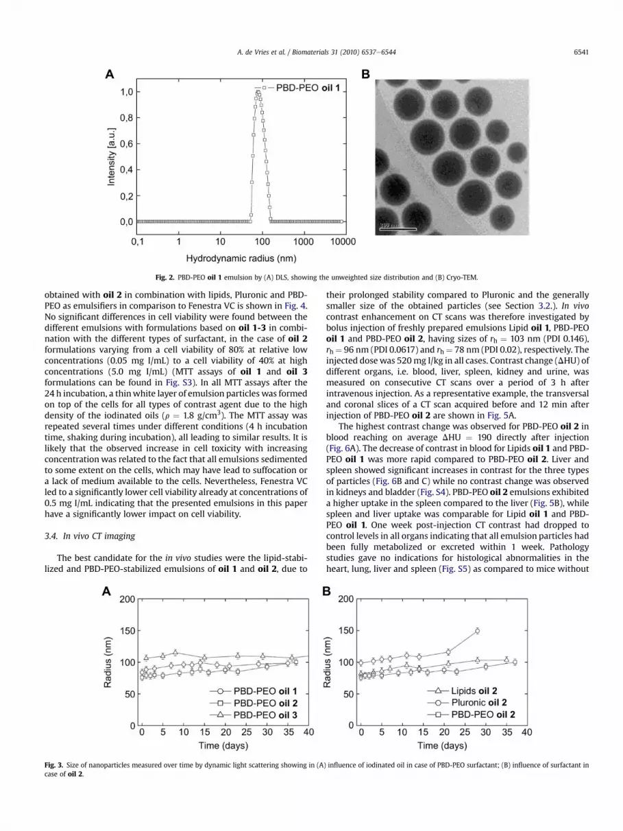
A typical size distribution of the PBD-PEO oil 1 emulsionobtained with dynamic light scattering (DLS) is presented in Fig. 2Ashowing a monomodal size distribution with a mean hydrody-namic radius of rh ¼ 84 nm (see Fig. S1 for number weighted data).PBD-PEO oil 1 filled emulsion particles are characterized by Cryo-TEM (Fig. 2B). All particles have a radius < 100 nm, typical for allsamples (Fig. S2) and have an electron dense core correlating withthe high iodine payload.
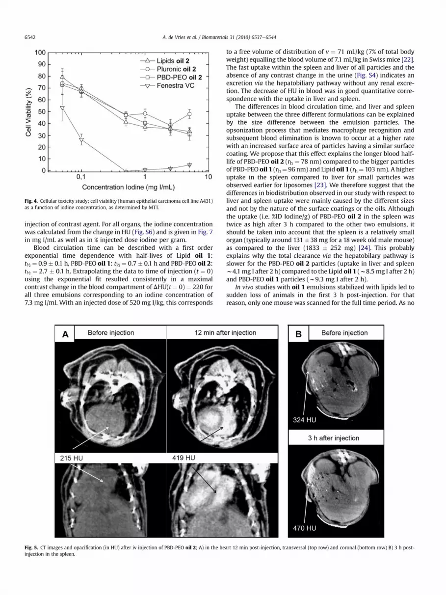
The mean hydrodynamic radius of the PBD-PEO emulsions of oil1, oil 2, and oil 3 respectively, was measured over time with DLS(Fig. 3A). A different hydrodynamic radius with rh(oil2) ¼ 76 nm < rh(oil 1) ¼ 84 nm < rh(oil 3) ¼ 101 nmwas obtainedshortly after preparation under identical processing conditions(Table 1). The hydrodynamic radius of these samples remainedessentially constant over a period of at least a month (Fig. 3A).
The mean hydrodynamic radius of the emulsions of oil 2,stabilized with lipids, Pluronic or PBD-PEO are measured over timewith DLS (Fig. 3B). A significant difference in radius was observedwhen using different surfactants i.e. for PBD-PEO: rh ¼ 76 � 4 nm,for Pluronic: rh ¼ 99� 4 nm, while in case of lipids: rh ¼ 82� 4 nm.Emulsions formulated with lipids or PBD-PEO yielded a stablemean hydrodynamic radius over time, while the mean size ofemulsions stabilizedwith Pluronic steadily increasedwith time andeven fell apart in two layers after day 28. Pluronic-stabilizedemulsions were therefore not considered for any in vivo studies.
A comparison between the particles prepared with different oilsand the calculated log P showed that smaller particles are obtainedwith the more hydrophobic oil 2 (log P ¼ 9.50) when compared toless hydrophobic oils (oil 1: log P ¼ 8.74; oil 3: log P ¼ 8.65). Asimilar relation between the log P of iodinated oil and the nano-particle size can be observed for lipid-stabilized and Pluronic-stabilized emulsion droplets (Table 1). The hydrophobicity of theiodinated oil seems therefore of eminent importance to obtainsmaller and therefore longer circulating emulsion particles.
The difference in radius observed for the three different emul-sifiers when using the same oils can be explained by their differentgeometrical constraints imposed by the size ratio of the hydrophilicto hydrophobic part. For an amphiphilic molecule the ratio of themolecular weight of the hydrophilic part over the total molecularweight (feo) can be used to describe this geometrical constraint. Forthe PBD-PEO the total molecular weight of the hydrophilic part ofthe ethylene oxide is larger than the hydrophobic block (feo ¼ 0.61;Mw,phil ¼ 2033 g/mol; Mw,phob ¼ 1305 g/mol) inducing a highersurface curvature in the particles. The PEO part likely extends intothe solution as surface grafted brush. Regular lipids self-assemblein more planar bilayers as the hydrophilic head group is smallerthan the hydrophobic part (feo ¼ 0.39; Mw,phil ¼ 312 g/mol; Mw,
phob¼ 478 g/mol). The formulation used here contains 5% pegylatedlipids (feow0.8), where the PEG molecules take a mushroom typeconfiguration at the surface but do not induce higher curvature. It ismore difficult to apply similar considerations to the triblock poly-mer Pluronic (feo ¼ 0.8, Mw,PEO ¼ 3360 g/mol; Mw,PPO¼ 1680 g/mol;Mw,PEO ¼ 3360 g/mol). We suspect that the triblock is less efficientto stabilize more strongly curved surfaces leading to the largestparticles compared to PBD-PEO and lipid stabilizers. Overall, thiswork shows that the hydrodynamic radius can be tuned with thechemical nature of the emulsifier and hydrophobic oil.
3.3. Cell viability
Acute cell toxicity of all emulsions was tested using a MTT assayand compared to the commercially available preclinical CT contrastagent Fenestra VC as a benchmark. The cell viability results
Fig. 2. PBD-PEO oil 1 emulsion by (A) DLS, showing the unweighted size distribution and (B) Cryo-TEM.
A. de Vries et al. / Biomaterials 31 (2010) 6537e6544 6541
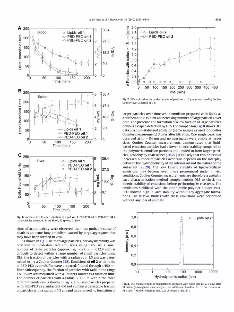
obtained with oil 2 in combination with lipids, Pluronic and PBD-PEO as emulsifiers in comparison to Fenestra VC is shown in Fig. 4.No significant differences in cell viability were found between thedifferent emulsions with formulations based on oil 1-3 in combi-nation with the different types of surfactant, in the case of oil 2formulations varying from a cell viability of 80% at relative lowconcentrations (0.05 mg I/mL) to a cell viability of 40% at highconcentrations (5.0 mg I/mL) (MTT assays of oil 1 and oil 3formulations can be found in Fig. S3). In all MTT assays after the24 h incubation, a thinwhite layer of emulsion particles was formedon top of the cells for all types of contrast agent due to the highdensity of the iodinated oils (r ¼ 1.8 g/cm3). The MTT assay wasrepeated several times under different conditions (4 h incubationtime, shaking during incubation), all leading to similar results. It islikely that the observed increase in cell toxicity with increasingconcentrationwas related to the fact that all emulsions sedimentedto some extent on the cells, which may have lead to suffocation ora lack of medium available to the cells. Nevertheless, Fenestra VCled to a significantly lower cell viability already at concentrations of0.5 mg I/mL indicating that the presented emulsions in this paperhave a significantly lower impact on cell viability.
3.4. In vivo CT imaging
The best candidate for the in vivo studies were the lipid-stabi-lized and PBD-PEO-stabilized emulsions of oil 1 and oil 2, due to
Fig. 3. Size of nanoparticles measured over time by dynamic light scattering showing in (A)case of oil 2.
their prolonged stability compared to Pluronic and the generallysmaller size of the obtained particles (see Section 3.2.). In vivocontrast enhancement on CT scans was therefore investigated bybolus injection of freshly prepared emulsions Lipid oil 1, PBD-PEOoil 1 and PBD-PEO oil 2, having sizes of rh ¼ 103 nm (PDI 0.146),rh¼ 96 nm (PDI 0.0617) and rh¼ 78 nm (PDI 0.02), respectively. Theinjected dosewas 520mg I/kg in all cases. Contrast change (DHU) ofdifferent organs, i.e. blood, liver, spleen, kidney and urine, wasmeasured on consecutive CT scans over a period of 3 h afterintravenous injection. As a representative example, the transversaland coronal slices of a CT scan acquired before and 12 min afterinjection of PBD-PEO oil 2 are shown in Fig. 5A.
The highest contrast change was observed for PBD-PEO oil 2 inblood reaching on average DHU ¼ 190 directly after injection(Fig. 6A). The decrease of contrast in blood for Lipids oil 1 and PBD-PEO oil 1 was more rapid compared to PBD-PEO oil 2. Liver andspleen showed significant increases in contrast for the three typesof particles (Fig. 6B and C) while no contrast change was observedin kidneys and bladder (Fig. S4). PBD-PEO oil 2 emulsions exhibiteda higher uptake in the spleen compared to the liver (Fig. 5B), whilespleen and liver uptake was comparable for Lipid oil 1 and PBD-PEO oil 1. One week post-injection CT contrast had dropped tocontrol levels in all organs indicating that all emulsion particles hadbeen fully metabolized or excreted within 1 week. Pathologystudies gave no indications for histological abnormalities in theheart, lung, liver and spleen (Fig. S5) as compared to mice without
influence of iodinated oil in case of PBD-PEO surfactant; (B) influence of surfactant in
Fig. 4. Cellular toxicity study; cell viability (human epithelial carcinoma cell line A431)as a function of iodine concentration, as determined by MTT.
A. de Vries et al. / Biomaterials 31 (2010) 6537e65446542
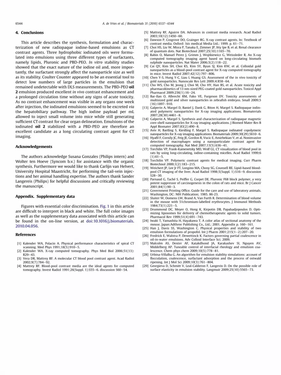
injection of contrast agent. For all organs, the iodine concentrationwas calculated from the change in HU (Fig. S6) and is given in Fig. 7in mg I/mL as well as in % injected dose iodine per gram.
Blood circulation time can be described with a first orderexponential time dependence with half-lives of Lipid oil 1:t½ ¼ 0.9 � 0.1 h, PBD-PEO oil 1: t½ ¼ 0.7 � 0.1 h and PBD-PEO oil 2:t½ ¼ 2.7 � 0.1 h. Extrapolating the data to time of injection (t ¼ 0)using the exponential fit resulted consistently in a maximalcontrast change in the blood compartment of DHU(t ¼ 0) ¼ 220 forall three emulsions corresponding to an iodine concentration of7.3 mg I/ml. With an injected dose of 520 mg I/kg, this corresponds
Fig. 5. CT images and opacification (in HU) after iv injection of PBD-PEO oil 2; A) in the heinjection in the spleen.
to a free volume of distribution of v ¼ 71 mL/kg (7% of total bodyweight) equalling the blood volume of 7.1 mL/kg in Swiss mice [22].The fast uptake within the spleen and liver of all particles and theabsence of any contrast change in the urine (Fig. S4) indicates anexcretion via the hepatobiliary pathway without any renal excre-tion. The decrease of HU in blood was in good quantitative corre-spondence with the uptake in liver and spleen.
The differences in blood circulation time, and liver and spleenuptake between the three different formulations can be explainedby the size difference between the emulsion particles. Theopsonization process that mediates macrophage recognition andsubsequent blood elimination is known to occur at a higher ratewith an increased surface area of particles having a similar surfacecoating. We propose that this effect explains the longer blood half-life of PBD-PEO oil 2 (rh ¼ 78 nm) compared to the bigger particlesof PBD-PEO oil 1 (rh¼ 96 nm) and Lipid oil 1 (rh¼ 103 nm). A higheruptake in the spleen compared to liver for small particles wasobserved earlier for liposomes [23]. We therefore suggest that thedifferences in biodistribution observed in our study with respect toliver and spleen uptake were mainly caused by the different sizesand not by the nature of the surface coatings or the oils. Althoughthe uptake (i.e. %ID Iodine/g) of PBD-PEO oil 2 in the spleen wastwice as high after 3 h compared to the other two emulsions, itshould be taken into account that the spleen is a relatively smallorgan (typically around 131�38mg for a 18 week old male mouse)as compared to the liver (1833 � 252 mg) [24]. This probablyexplains why the total clearance via the hepatobilary pathway isslower for the PBD-PEO oil 2 particles (uptake in liver and spleenw4.1mg I after 2 h) compared to the Lipid oil 1 (w8.5mg I after 2 h)and PBD-PEO oil 1 particles (w9.3 mg I after 2 h).
In vivo studies with oil 1 emulsions stabilized with lipids led tosudden loss of animals in the first 3 h post-injection. For thatreason, only one mouse was scanned for the full time period. As no
art 12 min post-injection, transversal (top row) and coronal (bottom row) B) 3 h post-
Fig. 6. Increase in HU after injection of Lipid oil 1, PBD-PEO oil 1, PBD-PEO oil 2nanoparticles measured in A) Blood, B) Spleen, C) Liver.
Fig. 7. Effect of surfactant on the number of particles > 1.5 mm as measured by CoulterCounter over a period of 7 h.
Fig. 8. DLS measurement of nanoparticles prepared with lipids and oil 2, 3 days afterfiltration (unweighted data analysis, no additional baseline fit to the correlationfunction) (number weighted data can be found in Fig. S7).
A. de Vries et al. / Biomaterials 31 (2010) 6537e6544 6543
signs of acute toxicity were observed, the most probable cause ofdeath is an acute lung embolism caused by large aggregates thatmay have been formed in vivo.
As shown in Fig. 3, neither large particles, nor any instability wasobserved in lipid-stabilized emulsions using DLS. As a smallnumber of large particles (approx.: rh > 2l; l ¼ 632.8 nm) isdifficult to detect within a large number of small particles usingDLS, the fraction of particles with a radius rh > 1.5 mm was deter-mined using a Coulter Counter [25]. Emulsions of oil 2 with lipids,or PBD-PEO as emulsifier were prepared, filtered through a 450 nmfilter. Subsequently, the fraction of particles with radii in the range1.5e15 mmwasmeasured with a Coulter Counter as a function time.The number of particles with a radius > 1.5 mm within the threedifferent emulsions is shown in Fig. 7. Emulsion particles preparedwith PBD-PEO as a surfactant did not contain a detectable fractionof particles with a radius> 1.5 mm and also showed no formation of
larger particles over time while emulsion prepared with lipids asa surfactant did exhibit an increasing number of large particles overtime. The presence and formation of a low fraction of large particlesobvious escaped detection by DLS. For comparison, Fig. 8 shows DLSdata of a lipid-stabilized emulsion (same sample as used for CoulterCounter measurement) 3 days after filtration. One single peak wasobserved at rh ¼ 84 nm and no aggregates were visible at largersizes. Coulter Counter measurements demonstrated that lipid-based emulsion particles had a lower kinetic stability compared tothe polymeric emulsion particles and tended to form larger parti-cles, probably by coalescence [26,27]. It is likely that this process ofincreased number of particles over time depends on the interplaybetween the hydrophobicity of the interior oil and the nature of thesurfactant [28,29]. The low kinetic stability of lipid-stabilizedemulsions may become even more pronounced under in vivoconditions. Coulter Counter measurements are therefore a useful invitro characterization method complementing DLS to check thekinetic stability of emulsions before performing in vivo tests. Theemulsions stabilized with the amphiphilic polymer diblock PBD-PEO showed high in vitro stability without any aggregate forma-tions. The in vivo studies with these emulsions were performedwithout any loss of animals.
A. de Vries et al. / Biomaterials 31 (2010) 6537e65446544
4. Conclusions
This article describes the synthesis, formulation and charac-terization of new radiopaque iodine-based emulsions as CTcontrast agents. Three hydrophobic iodinated oils were formu-lated into emulsions using three different types of surfactants,namely lipids, Pluronic and PBD-PEO. In vitro stability studiesshowed that the exact nature of the iodine oil and, more impor-tantly, the surfactant strongly affect the nanoparticle size as wellas its stability. Coulter Counter appeared to be an essential tool todetect low numbers of large particles in the emulsion thatremained undetectable with DLS measurements. The PBD-PEO oil2 emulsion produced excellent in vivo contrast enhancement anda prolonged circulation time without any signs of acute toxicity.As no contrast enhancement was visible in any organs one weekafter injection, the iodinated emulsions seemed to be excreted viathe hepatobiliary pathway. The high iodine payload per mLallowed to inject small volume into mice while still generatingsufficient CT contrast for clear organ delineation. Emulsions of theiodinated oil 2 stabilized with a PBD-PEO are therefore anexcellent candidate as a long circulating contrast agent for CTimaging.
Acknowledgements
The authors acknowledge Susana Gonzales (Philips intern) andWolter ten Hoeve (Syncom b.v.) for assistance with the organicsynthesis. Furthermore we would like to thank Carlijn van Helvert,University Hospital Maastricht, for performing the tail-vein injec-tions and her animal handling expertise. The authors thank SanderLangereis (Philips) for helpful discussions and critically reviewingthe manuscript.
Appendix. Supplementary data
Figures with essential color discrimination. Fig. 1 in this articleare difficult to interpret in black and white. The full color imagesas well as the supplementary data associated with this article canbe found in the on-line version, at doi:10.1016/j.biomaterials.2010.04.056.
References
[1] Kalender WA, Polacin A. Physical performance characteristics of spiral CTscanning. Med Phys 1991;18(5):910e5.
[2] Kalender WA. X-ray computed tomography. Phys Med Biol 2006;51(13):R29e43.
[3] Vera DR, Mattrey RF. A molecular CT blood pool contrast agent. Acad Radiol2002;9(7):784e92.
[4] Mattrey RF. Blood-pool contrast media are the ideal agents for computedtomography. Invest Radiol 1991;26(Suppl. 1):S55e6. discussion S60e54.
[5] Mattrey RF, Aguirre DA. Advances in contrast media research. Acad Radiol2003;10(12):1450e60.
[6] Dawson P, Cosgrove DO, Grainger RG. X-ray contrast agents. In: Textbook ofcontrast media. Oxford: Isis medical Media Ltd.; 1999. p. 61e71.
[7] Choi HS, Liu W, Misra P, Tanaka E, Zimmer JP, Itty Ipe B, et al. Renal clearanceof quantum dots. Nat Biotechnol 2007;25(10):1165e70.
[8] Rabin O, Manuel Perez J, Grimm J, Wojtkiewicz G, Weissleder R. An X-raycomputed tomography imaging agent based on long-circulating bismuthsulphide nanoparticles. Nat Mater 2006;5(2):118e22.
[9] Cai QY, Kim SH, Choi KS, Kim SY, Byun SJ, Kim KW, et al. Colloidal goldnanoparticles as a blood-pool contrast agent for X-ray computed tomographyin mice. Invest Radiol 2007;42(12):797e806.
[10] Chen Y-S, Hung Y-C, Liau I, Huang GS. Assessment of the in vivo toxicity ofgold nanoparticles. Nanoscale Res Lett 2009;4:858e64.
[11] Cho WS, Cho M, Jeong J, Choi M, Cho HY, Han BS, et al. Acute toxicity andpharmacokinetics of 13 nm-sized PEG-coated gold nanoparticles. Toxicol ApplPharmacol 2009;236(1):16e24.
[12] Bar-Ilan O, Albrecht RM, Fako VE, Furgeson DY. Toxicity assessments ofmultisized gold and silver nanoparticles in zebrafish embryos. Small 2009;5(16):1897e910.
[13] Galperin A, Margel D, Baniel J, Dank G, Biton H, Margel S. Radiopaque iodin-ated polymeric nanoparticles for X-ray imaging applications. Biomaterials2007;28(30):4461e8.
[14] Galperin A, Margel S. Synthesis and characterization of radiopaque magneticcore-shell nanoparticles for X-ray imaging applications. J Biomed Mater Res BAppl Biomater 2007;83(2):490e8.
[15] Aviv H, Bartling S, Kieslling F, Margel S. Radiopaque iodinated copolymericnanoparticles for X-ray imaging applications. Biomaterials 2009;30(29):5610e6.
[16] Hyafil F, Cornily JC, Feig JE, Gordon R, Vucic E, Amirbekian V, et al. Noninvasivedetection of macrophages using a nanoparticulate contrast agent forcomputed tomography. Nat Med 2007;13(5):636e41.
[17] Torchilin VP, Frank-Kamenetsky MD, Wolf GL. CT visualization of blood pool inrats by using long-circulating, iodine-containing micelles. Acad Radiol 1999;6(1):61e5.
[18] Torchilin VP. Polymeric contrast agents for medical imaging. Curr PharmBiotechnol 2000;1(2):183e215.
[19] Weichert JP, Lee Jr FT, Longino MA, Chosy SG, Counsell RE. Lipid-based blood-pool CT imaging of the liver. Acad Radiol 1998;5(Suppl. 1):S16e9. discussionS28e30.
[20] Parnaud G, Taché S, Peiffer G, Corpet DE. Pluronic F68 block polymer, a verypotent suppressor of carcinogenesis in the colon of rats and mice. Br J Cancer2001;84(1):90e3.
[21] Government Printing Office. Guide for the care and use of laboratory animals.Washington, DC: NIH Publication; 1985. 86-23.
[22] Sluiter W, Oomens LW, Brand A, Van Furth R. Determination of blood volumein the mouse with 51chromium-labelled erythrocytes. J Immunol Methods1984;73(1):221e5.
[23] Drummond DC, Meyer O, Hong K, Kirpotin DB, Papahadjopoulos D. Opti-mizing liposomes for delivery of chemotherapeutic agents to solid tumors.Pharmacol Rev 1999;51(4):691e743.
[24] Iwaki T, Yamashita H, Hayakawa T. A color atlas of sectional anatomy of themouse. Japan:Adthree Publishing Co., Ltd.; 2001. Appendix p. 160e161.
[25] Han J, Davis SS, Washington C. Physical properties and stability of twoemulsion formulations of propofol. Int J Pharm 2001;215(1e2):207e20.
[26] Fredrick E, Walstra P, Dewettinck K. Factors governing partial coalescence inoil-in-water emulsions. Adv Colloid Interface Sci; 2009.
[27] Malcolm AS, Dexter AF, Katakdhond JA, Karakashev SI, Nguyen AV,Middelberg AP. Tuneable control of interfacial rheology and emulsion coa-lescence. Chem phys chem 2009;10(5):778e81.
[28] Urbina-Villalba G. An algorithm for emulsion stability simulations: account offlocculation, coalescence, surfactant adsorption and the process of ostwaldripening. Int J Mol Sci 2009;10(3):761e804.
[29] Georgieva D, Schmitt V, Leal-Calderon F, Langevin D. On the possible role ofsurface elasticity in emulsion stability. Langmuir 2009;25(10):5565e73.


![[Kidney and iodinated and gadolinium-based contrast agents]](https://static.documents.page/doc/80x56/634e8a9e3bdc8e881007f2b5/kidney-and-iodinated-and-gadolinium-based-contrast-agents.jpg)


