+ All documents
Home > Documents > Olfactory Proteins Mediating Chemical Communication in the Navel Orangeworm Moth, Amyelois...

Olfactory Proteins Mediating Chemical Communication in the Navel Orangeworm Moth, Amyelois...

Date post: 10-Nov-2023
Category:
Upload: ucdavis
View: 0 times
Download: 0 times
Share this document with a friend
11
Olfactory Proteins Mediating Chemical Communication in the Navel Orangeworm Moth, Amyelois transitella Walter S. Leal 1 *, Yuko Ishida 1 , Julien Pelletier 1 , Wei Xu 1 , Josep Rayo 1 , Xianzhong Xu 2 , James B. Ames 2 1 Department of Entomology, University of California Davis, Davis, California, United States of America, 2 Department of Chemistry, University of California Davis, Davis, California, United States of America Abstract Background: The navel orangeworm, Amyelois transitella Walker (Lepidoptera: Pyralidae), is the most serious insect pest of almonds and pistachios in California for which environmentally friendly alternative methods of control — like pheromone- based approaches — are highly desirable. Some constituents of the sex pheromone are unstable and could be replaced with parapheromones, which may be designed on the basis of molecular interaction of pheromones and pheromone- detecting olfactory proteins. Methodology: By analyzing extracts from olfactory and non-olfactory tissues, we identified putative olfactory proteins, obtained their N-terminal amino acid sequences by Edman degradation, and used degenerate primers to clone the corresponding cDNAs by SMART RACE. Additionally, we used degenerate primers based on conserved sequences of known proteins to fish out other candidate olfactory genes. We expressed the gene encoding a newly identified pheromone- binding protein, which was analyzed by circular dichroism, fluorescence, and nuclear magnetic resonance, and used in a binding assay to assess affinity to pheromone components. Conclusion: We have cloned nine cDNAs encoding olfactory proteins from the navel orangeworm, including two pheromone-binding proteins, two general odorant-binding proteins, one chemosensory protein, one glutathione S- transferase, one antennal binding protein X, one sensory neuron membrane protein, and one odorant receptor. Of these, AtraPBP1 is highly enriched in male antennae. Fluorescence, CD and NMR studies suggest a dramatic pH-dependent conformational change, with high affinity to pheromone constituents at neutral pH and no binding at low pH. Citation: Leal WS, Ishida Y, Pelletier J, Xu W, Rayo J, et al. (2009) Olfactory Proteins Mediating Chemical Communication in the Navel Orangeworm Moth, Amyelois transitella. PLoS ONE 4(9): e7235. doi:10.1371/journal.pone.0007235 Editor: Mark A. Frye, UCLA - Physiological Science, United States of America Received August 11, 2009; Accepted September 8, 2009; Published September 30, 2009 Copyright: ß 2009 Leal et al. This is an open-access article distributed under the terms of the Creative Commons Attribution License, which permits unrestricted use, distribution, and reproduction in any medium, provided the original author and source are credited. Funding: This work was supported in part by National Science Foundation (0918177 to W.S.L), the Almond Board of California, California Pistachio Research Board (to W.S.L) and the National Institute of Health (EY012347 to J.B.A), but the funders had no role in study design, data collection, and analysis, decision to publish, or preparation of the manuscript. Competing Interests: The authors have declared that no competing interests exist. * E-mail: [email protected] Introduction Insects are biosensors par excellence. They have developed a remarkable ability to detect with extreme sensitivity and selectivity small, hydrophobic molecules that convey essential information for their reproduction and survival. Female moths, for example, advertize their readiness to mate by releasing infinitesimal amounts of a species-specific sex pheromone bouquet, which is remotely detected by males with remarkable precision. Minute amounts of signal deters eavesdropping, but requires such a fine tuning that the male olfactory system may be considered a ‘‘gold standard’’ in olfaction. It has been estimated that males of the silkworm moths, for example, can detect one molecule of the pheromone bombykol [1]. Moreover, small modifications in pheromone molecules render them completely inactive, or at least a few order of magnitude less active [2]. There is growing evidence in the literature suggesting that pheromone-binding proteins (PBPs) contribute to the sensitivity and possibly the selectivity of the olfactory system. PBPs are part of a family of olfactory proteins, including odorant-binding proteins (OBPs) and chemo- sensory proteins (CSPs), postulated to be involved in uptake of odorants, transport through the sensillar lymph, and delivery to membrane-bound odorant receptors. A detailed mechanism has been proposed for a pheromone- binding protein of the silkworm moth, BmorPBP1, suggesting that a pH-dependent conformational change is involved in pheromone binding and release [3,4,5,6]. Indeed, structural biology studies showed that the C-terminal part of the protein forms an additional a-helix at low pH that competes with pheromone molecules for the binding pocket [7,8,9], thus enabling the delivery of the pheromone in acidic environment similar to that formed by the negatively charged dendrite surfaces of the olfactory receptor neurons [10]. Functional studies also showed that BmorPBP1, when co-expressed with pheromone receptor BmorOR1 in the empty neuron system of Drosophila, enhanced the response to the pheromone, indicating that OBPs contribute to the remarkable sensitivity of the insect’s olfactory system [11]. The navel orangeworm, Amyelois transitella Walker (Lepidoptera: Pyralidae), is the most serious insect pest of almonds and pistachios in California, and a major pest of a number of other crops, including walnuts and figs. The navel orangeworm is primarily controlled during the growing season with pyrethroids and insect PLoS ONE | www.plosone.org 1 September 2009 | Volume 4 | Issue 9 | e7235
Transcript

Olfactory Proteins Mediating Chemical Communicationin the Navel Orangeworm Moth, Amyelois transitellaWalter S. Leal1*, Yuko Ishida1, Julien Pelletier1, Wei Xu1, Josep Rayo1, Xianzhong Xu2, James B. Ames2

1 Department of Entomology, University of California Davis, Davis, California, United States of America, 2 Department of Chemistry, University of California Davis, Davis,

California, United States of America

Abstract

Background: The navel orangeworm, Amyelois transitella Walker (Lepidoptera: Pyralidae), is the most serious insect pest ofalmonds and pistachios in California for which environmentally friendly alternative methods of control — like pheromone-based approaches — are highly desirable. Some constituents of the sex pheromone are unstable and could be replacedwith parapheromones, which may be designed on the basis of molecular interaction of pheromones and pheromone-detecting olfactory proteins.

Methodology: By analyzing extracts from olfactory and non-olfactory tissues, we identified putative olfactory proteins,obtained their N-terminal amino acid sequences by Edman degradation, and used degenerate primers to clone thecorresponding cDNAs by SMART RACE. Additionally, we used degenerate primers based on conserved sequences of knownproteins to fish out other candidate olfactory genes. We expressed the gene encoding a newly identified pheromone-binding protein, which was analyzed by circular dichroism, fluorescence, and nuclear magnetic resonance, and used in abinding assay to assess affinity to pheromone components.

Conclusion: We have cloned nine cDNAs encoding olfactory proteins from the navel orangeworm, including twopheromone-binding proteins, two general odorant-binding proteins, one chemosensory protein, one glutathione S-transferase, one antennal binding protein X, one sensory neuron membrane protein, and one odorant receptor. Of these,AtraPBP1 is highly enriched in male antennae. Fluorescence, CD and NMR studies suggest a dramatic pH-dependentconformational change, with high affinity to pheromone constituents at neutral pH and no binding at low pH.

Citation: Leal WS, Ishida Y, Pelletier J, Xu W, Rayo J, et al. (2009) Olfactory Proteins Mediating Chemical Communication in the Navel Orangeworm Moth, Amyeloistransitella. PLoS ONE 4(9): e7235. doi:10.1371/journal.pone.0007235

Editor: Mark A. Frye, UCLA - Physiological Science, United States of America

Received August 11, 2009; Accepted September 8, 2009; Published September 30, 2009

Copyright: � 2009 Leal et al. This is an open-access article distributed under the terms of the Creative Commons Attribution License, which permits unrestricteduse, distribution, and reproduction in any medium, provided the original author and source are credited.

Funding: This work was supported in part by National Science Foundation (0918177 to W.S.L), the Almond Board of California, California Pistachio ResearchBoard (to W.S.L) and the National Institute of Health (EY012347 to J.B.A), but the funders had no role in study design, data collection, and analysis, decision topublish, or preparation of the manuscript.

Competing Interests: The authors have declared that no competing interests exist.

* E-mail: [email protected]

Introduction

Insects are biosensors par excellence. They have developed a

remarkable ability to detect with extreme sensitivity and selectivity

small, hydrophobic molecules that convey essential information for

their reproduction and survival. Female moths, for example,

advertize their readiness to mate by releasing infinitesimal

amounts of a species-specific sex pheromone bouquet, which is

remotely detected by males with remarkable precision. Minute

amounts of signal deters eavesdropping, but requires such a fine

tuning that the male olfactory system may be considered a ‘‘gold

standard’’ in olfaction. It has been estimated that males of the

silkworm moths, for example, can detect one molecule of the

pheromone bombykol [1]. Moreover, small modifications in

pheromone molecules render them completely inactive, or at least

a few order of magnitude less active [2]. There is growing evidence

in the literature suggesting that pheromone-binding proteins

(PBPs) contribute to the sensitivity and possibly the selectivity of

the olfactory system. PBPs are part of a family of olfactory

proteins, including odorant-binding proteins (OBPs) and chemo-

sensory proteins (CSPs), postulated to be involved in uptake of

odorants, transport through the sensillar lymph, and delivery to

membrane-bound odorant receptors.

A detailed mechanism has been proposed for a pheromone-

binding protein of the silkworm moth, BmorPBP1, suggesting that

a pH-dependent conformational change is involved in pheromone

binding and release [3,4,5,6]. Indeed, structural biology studies

showed that the C-terminal part of the protein forms an additional

a-helix at low pH that competes with pheromone molecules for

the binding pocket [7,8,9], thus enabling the delivery of the

pheromone in acidic environment similar to that formed by the

negatively charged dendrite surfaces of the olfactory receptor

neurons [10]. Functional studies also showed that BmorPBP1,

when co-expressed with pheromone receptor BmorOR1 in the

empty neuron system of Drosophila, enhanced the response to the

pheromone, indicating that OBPs contribute to the remarkable

sensitivity of the insect’s olfactory system [11].

The navel orangeworm, Amyelois transitella Walker (Lepidoptera:

Pyralidae), is the most serious insect pest of almonds and pistachios

in California, and a major pest of a number of other crops,

including walnuts and figs. The navel orangeworm is primarily

controlled during the growing season with pyrethroids and insect

PLoS ONE | www.plosone.org 1 September 2009 | Volume 4 | Issue 9 | e7235

growth regulators, but alternative methods of control are sorely

needed. Sex pheromones offer an environmentally-friendly

alternative to control insect populations by mating disruption or

other strategies in integrated pest management. Typically, sex

pheromones and other attractants (aka semiochemicals) are

identified by a bioassay-guided isolation of natural products.

Alternatively, olfactory proteins may be used in a reverse chemical

ecology approach [12,13] for screening potential attractants on the

basis of their affinity to odorant-binding proteins. These proteins

are part of a large family of carrier proteins, for which we coined

the term encapsulins [12,14], but those directly involved in

semiochemical reception are grouped into pheromone-binding

proteins (PBPs) and general odorant-binding proteins (GOBPs)

based on their transport of pheromones or other semiochemicals

[15], respectively. We have now isolated, cloned and expressed

olfactory proteins from the navel orangeworm and set the stage to

use them in reverse chemical ecology. Although the sex

pheromone system of the navel orangeworm has already been

identified [16,17], some of the constituents are unstable. Reverse

chemical ecology in this case can be used for the development of

alternative compounds (parapheromones).

Results and Discussion

Isolation of antennae-specific proteinsTo isolate putative olfactory proteins from the navel orange-

worm, we extracted proteins from olfactory and non-olfactory

tissues dissected from adult males and females, and compared

protein profiles of these extracts by native polyacrylamide gel

electrophoresis (PAGE). Typically, OBPs are abundant acidic

proteins that migrate faster than non-olfactory proteins thus

appearing in the lower part of a native gel. Antennae-specific

proteins can be identified by comparing protein extracts from non-

olfactory tissues (e.g.: legs) with protein profiles from antennae. In

addition, comparison of male and female antennal extracts may

identify putative PBPs, which in most cases are specifically

expressed or at least enriched in male antennae. This protein-

based approach led us to identify several bands (Bands 1–7), which

are likely to represent olfactory proteins from the navel orange-

worm (Fig. 1). A faint band migrating just above Band 1 (Fig. 1)

was also detected in leg extracts when larger control samples were

analyzed (data not shown). To obtain the N-terminal amino acid

sequences of the target proteins by Edman degradation we re-ran

native PAGE analysis, transferred proteins to polyvinyl difluoride

membranes and isolated the bands after staining. Band 1 (Fig. 1)

was slightly more intense in extracts from female antennae, but

samples from male and female antennae gave the same N-terminal

sequence: SAEVMSHVTAHFGKA. Contrary to the initial

assumption that Bands 2 and 3 were different, they gave the

same N-terminal sequence: SQEVLHKMTASF. On the other

hand, Band 4 was detected exclusively in male antennae and gave

the N-terminal sequence SPEIMKDLSINFGKA. Bands 5 and 6

gave identical N-terminal sequences, DVAVMKDVTLGFGEA

with nearly equal intensity in extracts from male and female

antennae, whereas Band 7 (SDYKTGKIENINIQE) was detected

with higher intensity in male than female antennae.

Cloning of PBPs, GOBPs and a Chemosensory Protein(CSP)

Our PCR approach to clone the cDNAs encoding the isolated

proteins started with the isolation of total RNA from antennal

tissues and synthesis of first strand cDNA by the SMART RACE

cDNA Amplification. We used 39-RACE cDNA and 59-RACE

cDNA templates with degenerate primers, designed on the basis of

the identified N-terminal amino acid sequences, GCGT15 or

universal primer mix (UPM) primers and, subsequently, gene-

specific primers (GSPs). With primers designed on the basis of the

male-specific Band 4 we obtained a 711 bp-long cDNA encoding

164 amino acid residues, including 22 residues of a signal peptide,

which was assigned on the basis of the N-terminal sequence of the

mature protein. The mature protein contained the hallmark of

insect OBPs, six cysteine residues. Blastp search indicated that this

protein had 72, 70, and 69% identities with Synanthedon exitiosa PBP

(AAF06142) [18], Antheraea polyphemus PBP1 (CAA35592) [19] and

A. pernyi PBP2 (Q17078) [20]. Thus we named this protein

AtraPBP1 (Accession Number, GQ433364; calculated molecular

weight, 16,072 Da; pI, 4.99).

Next, we cloned the genes encoding the other antennae-specific

proteins starting with Band 1. Although we observed a rather

unusual connection of the SMART II Oligonucleotide (Clontech)

to the 59-end of the cDNA immediately after the 59-region

encoding the N-terminal amino acid sequence, we were able to

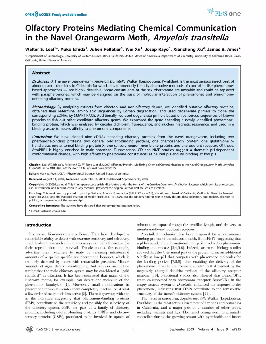
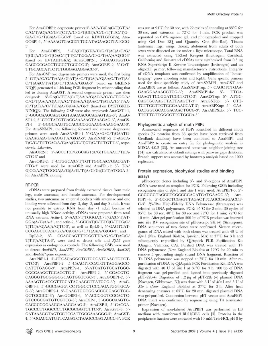
Figure 1. Analysis of proteins extracted from olfactory andnon-olfactory tissues. Protein extracts were separated on a 15%native PAGE and stained with Coomassie Brilliant Blue. Seven bandswere tentatively identified as antennae-specific. A band marked with adotted line, with slower migration than Band 1, was ruled out becausethe faint band in leg extracts was clearly detected in extracts from largesamples of this non-olfactory tissue. L, extract from male hindlegs (15legs-equivalent); MA, male antennae (70 antennae-equivalent); FA,female-antennae (65 antennae-equivalent).doi:10.1371/journal.pone.0007235.g001

Moth Olfactory Proteins

PLoS ONE | www.plosone.org 2 September 2009 | Volume 4 | Issue 9 | e7235

isolate a 511 bp-long cDNA encoding a 141-residue mature

protein with a N-terminal sequence identical to that of the isolated

protein. The translated protein had six cysteine residues and

showed 84, 81 and 79% identities with Manduca sexta GOBP2

(AAG50015) [21], A. pernyi GOBP2 (Q17075) [22], and Samia

cynthia ricini GOBP2 (BAF91328) (Leal, unpublished), respectively.

We, therefore, named the Band 1 protein AtraGOBP2 (Accession

Number, GQ433368; calculated molecular weight, 16,166 Da; pI,

4.87).

We had also encountered problems with 59-RACE when

cloning the cDNA encoding the protein in Bands 2 and 3, but

in this case the SMART II Oligonucleotide was connected

upstream of the 59-region encoding the N-terminal amino

sequence and the signal peptide. The cloned partial cDNA

sequence included 614 bp encoding for 21 amino acid residues of

the signal peptide and 146 residues of the mature protein. We

sequenced 16 independent clones and observed four points of

polymorphism at 138th C/G, 368th T/A, 521st A/T and 539th T/A,

which suggest the occurrence of two forms of the mature protein,

one with Phe-102 and the other having Tyr-102, and both having

six cysteine residues. Blastp search indicated 67, 66, and 65%

identity to pheromone-binding proteins from Heliothis virescens PBP2

(CAL48346) [23], Helicoverpa assulta PBP2 (ABY28381) (Yang, W. L.

et al., unpublished), and H. armigera PBP2 (ACD01993) (Zhang, S.

et al, unpublished), respectively. Therefore, we named the two

forms AtraPBP2F102 (Accession Number, GQ433365; calculated

molecular weight, 16,731 Da; pI, 5.15) and AtraPBP2Y102

(Accession Number, GQ433366; calculated molecular weight,

16,747 Da; pI, 5.15).

With degenerate primers for the protein in Bands 5 and 6, we

isolated a partial cDNA sequence of 702 bp encoding 144 amino

acid residues of the mature protein, which included seven cysteine

residues, and four residues of the signal peptide. Blastp search

indicated that the protein had 78, 77, and 76% identities with

GOBP1s from Bombyx mori (CAAA64444) [24], Plutella xylostella

(ABW05104) (Dong, X.-L. et al., unpublished) and H. virescens

(CAA65605) [25]. These data suggest that the protein in Bands 5

and 6 belongs to the seven-cysteine group of GOBP1s from moths.

Consequently, we named this protein AtraGOBP1 (Accession

Number, GQ433367; calculated molecular weight, 16,903 Da; pI,

5.11).

Lastly, we cloned the cDNA encoding the protein detected in

Band 7. A partial 537 bp-long cDNA was isolated, which encoded

for 105 amino acid residues, including the N-terminal sequence

obtained by Edman degradation. This protein was named

AtraCSP (Accession Number, GQ433369; calculated molecular

weight, 12,923 Da; pI, 5.63) because the mature protein contains

four cysteine residues and showed 80, 77, and 73% amino acid

identity to chemosensory proteins from B. mori (AAV34688) [26],

Cactoblastis cactoris (AAC47827) [27], and H. armigera (AF368375)

(Deyts et. al., unpublished), respectively.

Cloning of other olfactory proteinsWhile attempting to clone the cDNA encoding AtraCSP, we

isolated a cDNA fragment encoding a glutathione S-transferase.

Because these enzymes are implicated in odorant reception [28],

we have designed gene-specific primers, obtained the entire

sequence by 59-RACE, and named this protein AtraGST

(Accession Number, GQ433371).

Using a degenerate primer PCR approach, we have identified a

partial cDNA sequence encoding a putative sensory neuron

membrane protein (SNMP). AtraSNMP1 (942 bp, 314aa) (Acces-

sion Number, GQ451327) displays 75% amino-acid identity to

Mamestra brassicae SNMP1 (AF462066) (Jacquin-Joly, E. et al.,

unpublished) and H. virescens SNMP1 (AJ251959) and 74% to B.

mori SNMP1 (AJ251958) [29]. Likewise, we have identified a

partial cDNA sequence encoding a putative atypical OR83b-like

odorant receptor 2 (OR2). AtraOR2 (813 bp, 271aa) (GQ451328)

is highly conserved, sharing 85 to 91% amino-acid identity with

orthologs of other lepidopteran species. While attempting to clone

cDNAs encoding pheromone receptors using degenerate forward

primer and universal primer, we have identified a partial cDNA

sequence encoding a putative antennal binding protein X (ABPX).

AtraABPX (421 bp, 117aa) (Accession Number, GQ451326)

displays 69% amino-acid identity to H. virescens ABPX

(AJ002518) [30], 64% to Agrotis ipsilon ABPX (AY301981)

(Picimbon, J.-F. et al., unpublished) and 63% to B. mori ABPX

(X94990) [31].

Expression patterns of olfactory proteins andphylogenetic relationships

To compare transcript patterns with protein profiles (Fig. 1),

RT-PCR experiments were performed using gene-specific prim-

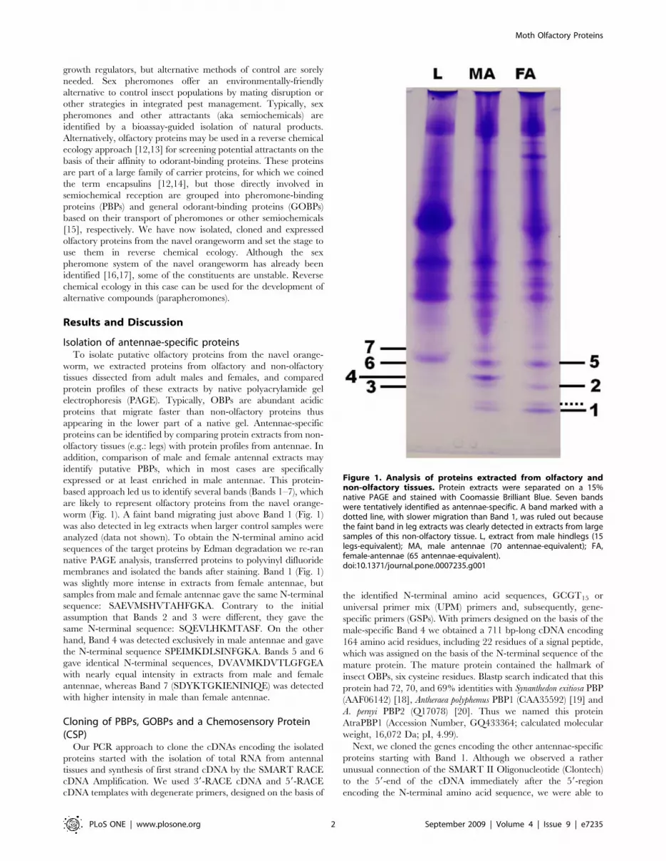
ers. First, we compared expression of AtraPBP1, AtraPBP2,

AtraGOBP1, AtraGOBP2, and AtraCSP in non-olfactory tissues

(male legs) with olfactory tissues (male and female antennae)

(Fig. 2). In general, gene expression mirrored protein profiles,

except for AtraCSP, which was detected not only in male and

female antennae, but also in non-olfactory tissues (legs). AtraPBP2,

AtraGOBP1 and AtraGOBP2 genes were detected in both male and

female antennae, but not in legs, whereas AtraPBP1 was apparently

expressed exclusively in male antennae. Next, we assessed gene

expression during antennal development. Contrary to our previous

experience with the wild silkworm moth, A. polyphemus [32],

sampling antennal pockets from pupae and day 0 adults of the

navel orangeworm and extracting RNA were very challenging due

to high RNAse activity at this developmental stage as reflected in

the irregular amplifications of actin control gene (Fig. 3). Indeed,

we were unable to extract RNA sample just the day before adult

eclosion (day -1). Despite the unavoidable fluctuation in template

titers, these experiments suggest that gene expression of most

olfactory proteins starts at least two days before adult emergence

(Fig. 3). Expression of the male antennae-specific AtraPBP1 starts

at day 0 of adult stage or the day prior to adult emergence.

Figure 2. Gene expression analysis by RT-PCR. Expression ofAtraPBP1, AtraPBP2, AtraGOBP1, AtraGOBP2, and AtraCSP genes incontrol tissue (ML, male hindlegs) and olfactory tissues (MA, maleantennae and FA, female antennae). Actin gene was used asendogenous control.doi:10.1371/journal.pone.0007235.g002

Moth Olfactory Proteins

PLoS ONE | www.plosone.org 3 September 2009 | Volume 4 | Issue 9 | e7235

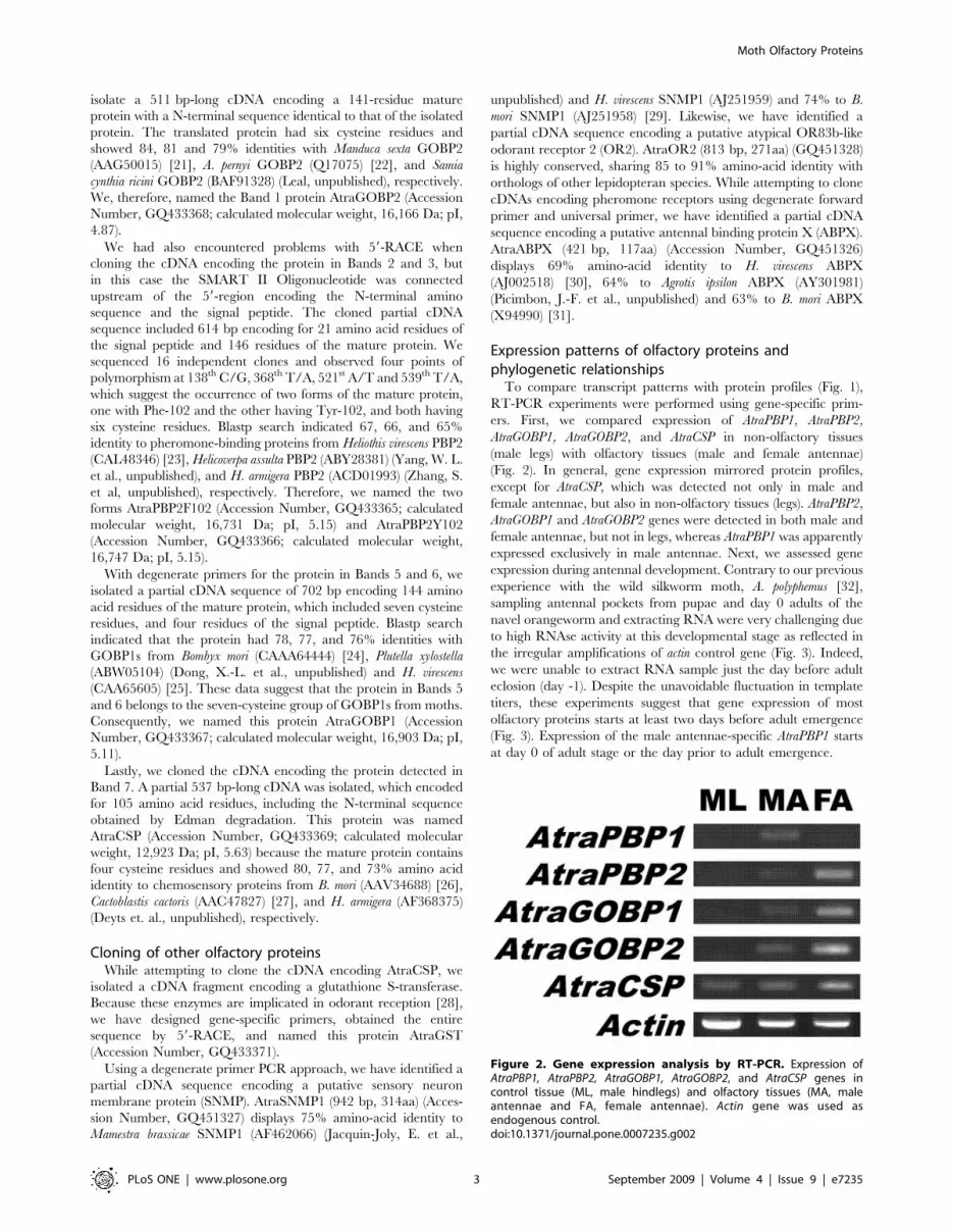
Having observed by non-quantitative RT-PCR that AtraPBP1

gene is expressed only in male antennae, a more thorough

examination of gene expression was performed. Indeed, AtraPBP1

was limited to expression in male antennae (Fig. 4), with no trace

detected in non-olfactory tissues, including legs, wings, thorax, and

abdomen. It is worth mentioning, however, that a faint band was

observed when cDNA from female antennae was used as template

thus suggesting that AtraPBP1 is highly enriched in male antennae.

Consequently, it is reasonable to assume that AtraPBP1 plays

male-specific role(s), such as the detection of sex pheromones.

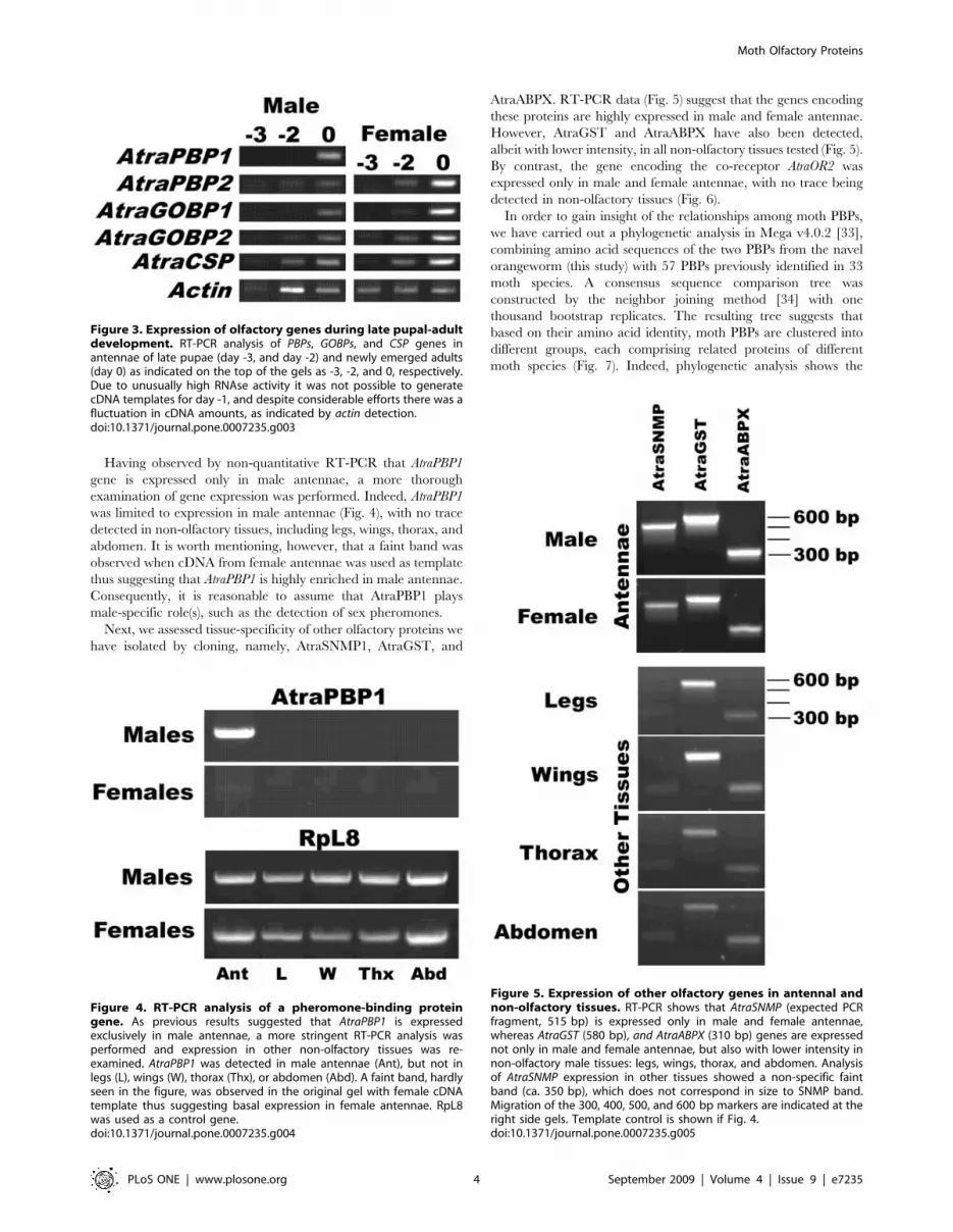
Next, we assessed tissue-specificity of other olfactory proteins we

have isolated by cloning, namely, AtraSNMP1, AtraGST, and

AtraABPX. RT-PCR data (Fig. 5) suggest that the genes encoding

these proteins are highly expressed in male and female antennae.

However, AtraGST and AtraABPX have also been detected,

albeit with lower intensity, in all non-olfactory tissues tested (Fig. 5).

By contrast, the gene encoding the co-receptor AtraOR2 was

expressed only in male and female antennae, with no trace being

detected in non-olfactory tissues (Fig. 6).

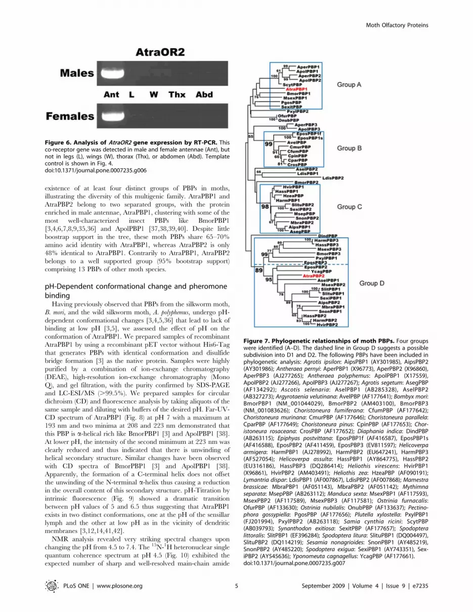
In order to gain insight of the relationships among moth PBPs,

we have carried out a phylogenetic analysis in Mega v4.0.2 [33],

combining amino acid sequences of the two PBPs from the navel

orangeworm (this study) with 57 PBPs previously identified in 33

moth species. A consensus sequence comparison tree was

constructed by the neighbor joining method [34] with one

thousand bootstrap replicates. The resulting tree suggests that

based on their amino acid identity, moth PBPs are clustered into

different groups, each comprising related proteins of different

moth species (Fig. 7). Indeed, phylogenetic analysis shows the

Figure 3. Expression of olfactory genes during late pupal-adultdevelopment. RT-PCR analysis of PBPs, GOBPs, and CSP genes inantennae of late pupae (day -3, and day -2) and newly emerged adults(day 0) as indicated on the top of the gels as -3, -2, and 0, respectively.Due to unusually high RNAse activity it was not possible to generatecDNA templates for day -1, and despite considerable efforts there was afluctuation in cDNA amounts, as indicated by actin detection.doi:10.1371/journal.pone.0007235.g003

Figure 4. RT-PCR analysis of a pheromone-binding proteingene. As previous results suggested that AtraPBP1 is expressedexclusively in male antennae, a more stringent RT-PCR analysis wasperformed and expression in other non-olfactory tissues was re-examined. AtraPBP1 was detected in male antennae (Ant), but not inlegs (L), wings (W), thorax (Thx), or abdomen (Abd). A faint band, hardlyseen in the figure, was observed in the original gel with female cDNAtemplate thus suggesting basal expression in female antennae. RpL8was used as a control gene.doi:10.1371/journal.pone.0007235.g004

Figure 5. Expression of other olfactory genes in antennal andnon-olfactory tissues. RT-PCR shows that AtraSNMP (expected PCRfragment, 515 bp) is expressed only in male and female antennae,whereas AtraGST (580 bp), and AtraABPX (310 bp) genes are expressednot only in male and female antennae, but also with lower intensity innon-olfactory male tissues: legs, wings, thorax, and abdomen. Analysisof AtraSNMP expression in other tissues showed a non-specific faintband (ca. 350 bp), which does not correspond in size to SNMP band.Migration of the 300, 400, 500, and 600 bp markers are indicated at theright side gels. Template control is shown if Fig. 4.doi:10.1371/journal.pone.0007235.g005

Moth Olfactory Proteins

PLoS ONE | www.plosone.org 4 September 2009 | Volume 4 | Issue 9 | e7235

existence of at least four distinct groups of PBPs in moths,

illustrating the diversity of this multigenic family. AtraPBP1 and

AtraPBP2 belong to two separated groups, with the protein

enriched in male antennae, AtraPBP1, clustering with some of the

most well-characterized insect PBPs like BmorPBP1

[3,4,6,7,8,9,35,36] and ApolPBP1 [37,38,39,40]. Despite little

boostrap support in the tree, these moth PBPs share 65–70%

amino acid identity with AtraPBP1, whereas AtraPBP2 is only

48% identical to AtraPBP1. Contrarily to AtraPBP1, AtraPBP2

belongs to a well supported group (95% bootstrap support)

comprising 13 PBPs of other moth species.

pH-Dependent conformational change and pheromonebinding

Having previously observed that PBPs from the silkworm moth,

B. mori, and the wild silkworm moth, A. polyphemus, undergo pH-

dependent conformational changes [3,4,5,36] that lead to lack of

binding at low pH [3,5], we assessed the effect of pH on the

conformation of AtraPBP1. We prepared samples of recombinant

AtraPBP1 by using a recombinant pET vector without His6-Tag

that generates PBPs with identical conformation and disulfide

bridge formation [3] as the native protein. Samples were highly

purified by a combination of ion-exchange chromatography

(DEAE), high-resolution ion-exchange chromatography (Mono

Q), and gel filtration, with the purity confirmed by SDS-PAGE

and LC-ESI/MS (.99.5%). We prepared samples for circular

dichroism (CD) and fluorescence analysis by taking aliquots of the

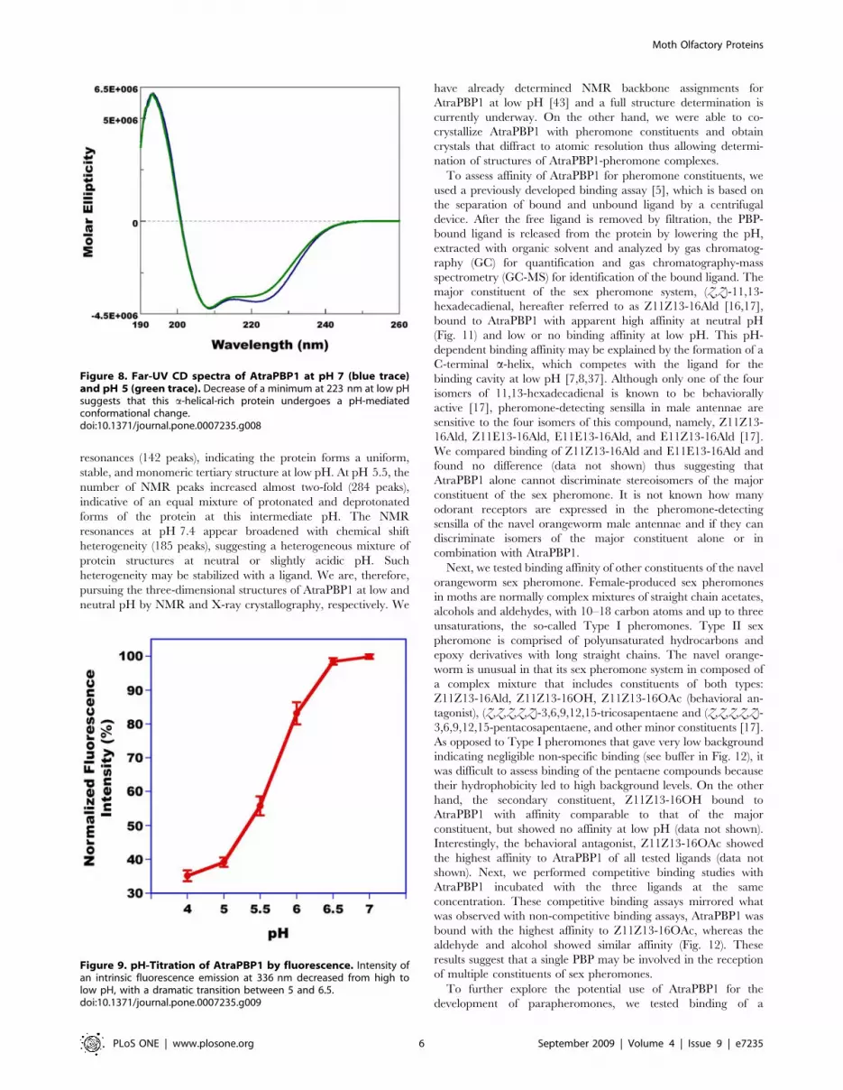
same sample and diluting with buffers of the desired pH. Far-UV-

CD spectrum of AtraPBP1 (Fig. 8) at pH 7 with a maximum at

193 nm and two minima at 208 and 223 nm demonstrated that

this PBP is a-helical rich like BmorPBP1 [3] and ApolPBP1 [38].

At lower pH, the intensity of the second minimum at 223 nm was

clearly reduced and thus indicated that there is unwinding of

helical secondary structure. Similar changes have been observed

with CD spectra of BmorPBP1 [3] and ApolPBP1 [38].

Apparently, the formation of a C-terminal helix does not offset

the unwinding of the N-terminal a-helix thus causing a reduction

in the overall content of this secondary structure. pH-Titration by

intrinsic fluorescence (Fig. 9) showed a dramatic transition

between pH values of 5 and 6.5 thus suggesting that AtraPBP1

exists in two distinct conformations, one at the pH of the sensillar

lymph and the other at low pH as in the vicinity of dendritic

membranes [3,12,14,41,42].

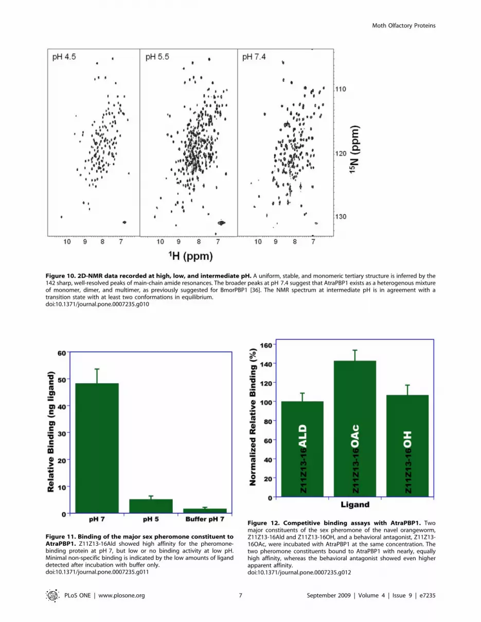
NMR analysis revealed very striking spectral changes upon

changing the pH from 4.5 to 7.4. The 15N-1H heteronuclear single

quantum coherence spectrum at pH 4.5 (Fig. 10) exhibited the

expected number of sharp and well-resolved main-chain amide

Figure 6. Analysis of AtraOR2 gene expression by RT-PCR. Thisco-receptor gene was detected in male and female antennae (Ant), butnot in legs (L), wings (W), thorax (Thx), or abdomen (Abd). Templatecontrol is shown in Fig. 4.doi:10.1371/journal.pone.0007235.g006

Figure 7. Phylogenetic relationships of moth PBPs. Four groupswere identified (A–D). The dashed line in Group D suggests a possiblesubdivision into D1 and D2. The following PBPs have been included inphylogenetic analysis: Agrotis ipsilon: AipsPBP1 (AY301985), AipsPBP2(AY301986); Antheraea pernyi: AperPBP1 (X96773), AperPBP2 (X96860),AperPBP3 (AJ277265); Antheraea polyphemus: ApolPBP1 (X17559),ApolPBP2 (AJ277266), ApolPBP3 (AJ277267); Agrotis segetum: AsegPBP(AF134292); Ascotis selenaria: AselPBP1 (AB285328), AselPBP2(AB327273); Argyrotaenia velutinana: AvelPBP (AF177641); Bombyx mori:BmorPBP1 (NM_001044029), BmorPBP2 (AM403100), BmorPBP3(NM_001083626); Choristoneura fumiferana: CfumPBP (AF177642);Choristoneura murinana: CmurPBP (AF177646); Choristoneura parallela:CparPBP (AF177649); Choristoneura pinus: CpinPBP (AF177653); Chor-istoneura rosaceana: CrosPBP (AF177652); Diaphania indica: DindPBP(AB263115); Epiphyas postvittana: EposPBP1f (AF416587), EposPBP1s(AF416588), EposPBP2 (AF411459), EposPBP3 (EV811597); Helicoverpaarmigera: HarmPBP1 (AJ278992), HarmPBP2 (EU647241), HarmPBP3(AF527054); Helicoverpa assulta: HassPBP1 (AY864775), HassPBP2(EU316186), HassPBP3 (DQ286414); Heliothis virescens: HvirPBP1(X96861), HvirPBP2 (AM403491); Heliothis zea: HzeaPBP (AF090191);Lymantria dispar: LdisPBP1 (AF007867), LdisPBP2 (AF007868); Mamestrabrassicae: MbraPBP1 (AF051143), MbraPBP2 (AF051142); Mythimnaseparata: MsepPBP (AB263112); Manduca sexta: MsexPBP1 (AF117593),MsexPBP2 (AF117589), MsexPBP3 (AF117581); Ostrinia furnacalis:OfurPBP (AF133630); Ostrinia nubilalis: OnubPBP (AF133637); Pectino-phora gossypiella: PgosPBP (AF177656); Plutella xylostella: PxylPBP1(FJ201994), PxylPBP2 (AB263118); Samia cynthia ricini: ScytPBP(AB039793); Synanthodon exitiosa: SexitPBP (AF177657); Spodopteralittoralis: SlitPBP1 (EF396284); Spodoptera litura: SlituPBP1 (DQ004497),SlituPBP2 (DQ114219); Sesamia nonagrioides: SnonPBP1 (AY485219),SnonPBP2 (AY485220); Spodoptera exigua: SexiPBP1 (AY743351), Sex-iPBP2 (AY545636); Yponomeuta cagnagellus: YcagPBP (AF177661).doi:10.1371/journal.pone.0007235.g007

Moth Olfactory Proteins

PLoS ONE | www.plosone.org 5 September 2009 | Volume 4 | Issue 9 | e7235

resonances (142 peaks), indicating the protein forms a uniform,

stable, and monomeric tertiary structure at low pH. At pH 5.5, the

number of NMR peaks increased almost two-fold (284 peaks),

indicative of an equal mixture of protonated and deprotonated

forms of the protein at this intermediate pH. The NMR

resonances at pH 7.4 appear broadened with chemical shift

heterogeneity (185 peaks), suggesting a heterogeneous mixture of

protein structures at neutral or slightly acidic pH. Such

heterogeneity may be stabilized with a ligand. We are, therefore,

pursuing the three-dimensional structures of AtraPBP1 at low and

neutral pH by NMR and X-ray crystallography, respectively. We

have already determined NMR backbone assignments for

AtraPBP1 at low pH [43] and a full structure determination is

currently underway. On the other hand, we were able to co-

crystallize AtraPBP1 with pheromone constituents and obtain

crystals that diffract to atomic resolution thus allowing determi-

nation of structures of AtraPBP1-pheromone complexes.

To assess affinity of AtraPBP1 for pheromone constituents, we

used a previously developed binding assay [5], which is based on

the separation of bound and unbound ligand by a centrifugal

device. After the free ligand is removed by filtration, the PBP-

bound ligand is released from the protein by lowering the pH,

extracted with organic solvent and analyzed by gas chromatog-

raphy (GC) for quantification and gas chromatography-mass

spectrometry (GC-MS) for identification of the bound ligand. The

major constituent of the sex pheromone system, (Z,Z)-11,13-

hexadecadienal, hereafter referred to as Z11Z13-16Ald [16,17],

bound to AtraPBP1 with apparent high affinity at neutral pH

(Fig. 11) and low or no binding affinity at low pH. This pH-

dependent binding affinity may be explained by the formation of a

C-terminal a-helix, which competes with the ligand for the

binding cavity at low pH [7,8,37]. Although only one of the four

isomers of 11,13-hexadecadienal is known to be behaviorally

active [17], pheromone-detecting sensilla in male antennae are

sensitive to the four isomers of this compound, namely, Z11Z13-

16Ald, Z11E13-16Ald, E11E13-16Ald, and E11Z13-16Ald [17].

We compared binding of Z11Z13-16Ald and E11E13-16Ald and

found no difference (data not shown) thus suggesting that

AtraPBP1 alone cannot discriminate stereoisomers of the major

constituent of the sex pheromone. It is not known how many

odorant receptors are expressed in the pheromone-detecting

sensilla of the navel orangeworm male antennae and if they can

discriminate isomers of the major constituent alone or in

combination with AtraPBP1.

Next, we tested binding affinity of other constituents of the navel

orangeworm sex pheromone. Female-produced sex pheromones

in moths are normally complex mixtures of straight chain acetates,

alcohols and aldehydes, with 10–18 carbon atoms and up to three

unsaturations, the so-called Type I pheromones. Type II sex

pheromone is comprised of polyunsaturated hydrocarbons and

epoxy derivatives with long straight chains. The navel orange-

worm is unusual in that its sex pheromone system in composed of

a complex mixture that includes constituents of both types:

Z11Z13-16Ald, Z11Z13-16OH, Z11Z13-16OAc (behavioral an-

tagonist), (Z,Z,Z,Z,Z)-3,6,9,12,15-tricosapentaene and (Z,Z,Z,Z,Z)-

3,6,9,12,15-pentacosapentaene, and other minor constituents [17].

As opposed to Type I pheromones that gave very low background

indicating negligible non-specific binding (see buffer in Fig. 12), it

was difficult to assess binding of the pentaene compounds because

their hydrophobicity led to high background levels. On the other

hand, the secondary constituent, Z11Z13-16OH bound to

AtraPBP1 with affinity comparable to that of the major

constituent, but showed no affinity at low pH (data not shown).

Interestingly, the behavioral antagonist, Z11Z13-16OAc showed

the highest affinity to AtraPBP1 of all tested ligands (data not

shown). Next, we performed competitive binding studies with

AtraPBP1 incubated with the three ligands at the same

concentration. These competitive binding assays mirrored what

was observed with non-competitive binding assays, AtraPBP1 was

bound with the highest affinity to Z11Z13-16OAc, whereas the

aldehyde and alcohol showed similar affinity (Fig. 12). These

results suggest that a single PBP may be involved in the reception

of multiple constituents of sex pheromones.

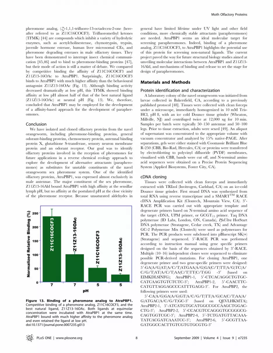
To further explore the potential use of AtraPBP1 for the

development of parapheromones, we tested binding of a

Figure 8. Far-UV CD spectra of AtraPBP1 at pH 7 (blue trace)and pH 5 (green trace). Decrease of a minimum at 223 nm at low pHsuggests that this a-helical-rich protein undergoes a pH-mediatedconformational change.doi:10.1371/journal.pone.0007235.g008

Figure 9. pH-Titration of AtraPBP1 by fluorescence. Intensity ofan intrinsic fluorescence emission at 336 nm decreased from high tolow pH, with a dramatic transition between 5 and 6.5.doi:10.1371/journal.pone.0007235.g009

Moth Olfactory Proteins

PLoS ONE | www.plosone.org 6 September 2009 | Volume 4 | Issue 9 | e7235

Figure 10. 2D-NMR data recorded at high, low, and intermediate pH. A uniform, stable, and monomeric tertiary structure is inferred by the142 sharp, well-resolved peaks of main-chain amide resonances. The broader peaks at pH 7.4 suggest that AtraPBP1 exists as a heterogenous mixtureof monomer, dimer, and multimer, as previously suggested for BmorPBP1 [36]. The NMR spectrum at intermediate pH is in agreement with atransition state with at least two conformations in equilibrium.doi:10.1371/journal.pone.0007235.g010

Figure 11. Binding of the major sex pheromone constituent toAtraPBP1. Z11Z13-16Ald showed high affinity for the pheromone-binding protein at pH 7, but low or no binding activity at low pH.Minimal non-specific binding is indicated by the low amounts of liganddetected after incubation with buffer only.doi:10.1371/journal.pone.0007235.g011

Figure 12. Competitive binding assays with AtraPBP1. Twomajor constituents of the sex pheromone of the navel orangeworm,Z11Z13-16Ald and Z11Z13-16OH, and a behavioral antagonist, Z11Z13-16OAc, were incubated with AtraPBP1 at the same concentration. Thetwo pheromone constituents bound to AtraPBP1 with nearly, equallyhigh affinity, whereas the behavioral antagonist showed even higherapparent affinity.doi:10.1371/journal.pone.0007235.g012

Moth Olfactory Proteins

PLoS ONE | www.plosone.org 7 September 2009 | Volume 4 | Issue 9 | e7235

pheromone analog, (Z)-1,1,1-trifluoro-13-octadecen-2-one (here-

after referred to as Z11C16COCF3). Trifluoromethyl ketones

(TFMK) [44] are compounds which inhibit a variety of hydrolytic

enzymes, such as acetylcholinesterase, chymotrypsin, trypsin,

juvenile hormone esterase, human liver microsomal CEs, and

pheromone degrading esterases in male olfactory tissues. They

have been demonstrated to interrupt insect chemical communi-

cation [45,46] and to bind to pheromone-binding proteins [47],

but their mode of action is still a matter of debate. We compared

by competitive binding the affinity of Z11C16COCF3 and

Z11Z13-16OAc to AtraPBP1. Surprisingly, Z11C16COCF3

binds to AtraPBP1 with much higher affinity than the behavioural

antagonist Z11Z13-16OAc (Fig. 13). Although binding activity

decreased dramatically at low pH, this TFMK showed binding

affinity at low pH almost half of that of the best natural ligand

(Z11Z13-16OAc) at neutral pH (Fig. 13). We, therefore,

concluded that AtraPBP1 may be employed for the development

of a affinity-based approach for the development of parapher-

omones.

ConclusionWe have isolated and cloned olfactory proteins from the navel

orangeworm, including pheromone-binding proteins, general

odorant-binding proteins, chemosensory protein, antennal binding

protein X, glutathione S-transferase, sensory neuron membrane

protein and an odorant receptor. Our goal was to identify

olfactory proteins involved in the reception of pheromones for

future applications in a reverse chemical ecology approach to

explore the development of alternative attractants (paraphero-

mones) as substitutes for unstable constituents of the navel

orangeworm sex pheromone system. One of the identified

olfactory proteins, AtraPBP1, was expressed almost exclusively in

male antennae. The major constituent of the sex pheromone,

Z11Z13-16Ald bound AtraPBP1 with high affinity at the sensillar

lymph pH, but no affinity at the postulated pH at the close vicinity

of the pheromone receptor. Because unsaturated aldehydes in

general have limited lifetime under UV light and other field

conditions, more chemically stable attractants (parapheromones)

are needed. AtraPBP1 seems an ideal molecular target for

screening parapheromones. Indeed, binding of a pheromone

analog, Z11C16COCF3, to AtraPBP1 highlights the potential use

of this protein for screening non-natural ligands. The current

project paved the way for future structural biology studies aimed at

unveiling molecular interactions between AtraPBP1 and Z11Z13-

16Ald, and mechanisms of binding and release to set the stage for

design of parapheromones.

Materials and Methods

Protein identification and characterizationA laboratory colony of the navel orangeworm was initiated from

larvae collected in Bakersfield, CA, according to a previously

published protocol [48]. Tissues were collected with clean forceps

under a microscope, immediately homogenized in 10 mM Tris-

HCl, pH 8, with an ice cold Dounce tissue grinder (Wheaton,

Millville, NJ) and centrifuged twice at 12,000 xg for 10 min.

Samples per batch were typically 50–150 antennae and 50–100

legs. Prior to tissue extraction, adults were sexed [49]. An aliquot

of supernatant was concentrated to the appropriate volume with

vacuum concentrator and analyzed by 15% native-PAGE. After

separations, gels were either stained with Coomassie Brilliant Blue

R-250 (CBB, Bio-Rad, Hercules, CA) or proteins were transferred

by electroblotting to polyvinyl difluoride (PVDF) membranes,

visualized with CBB, bands were cut off, and N-terminal amino

acid sequences were obtained on a Precise Protein Sequencing

System (Applied Biosystems, Foster City, CA).

cDNA cloningTissues were collected with clean forceps and immediately

extracted with TRIzol (Invitrogen, Carlsbad, CA) on an ice-cold

Dounce tissue grinder. First strand DNA was synthesized from

total RNA using reverse transcriptase and a SMARTTM RACE

cDNA Amplification Kit (Clontech, Mountain View, CA). 39-

RACE PCR was carried out with appropriate template and

degenerate primers based on N-terminal amino acid sequence of

the target cDNA, UPM primer, or GCGT15 primer. Taq DNA

polymerase (ID Labs, London, ON, Canada), PfuUltra HotStart

DNA polymerase (Stratagene, Cedar creek, TX) and Advantage

GC-2 Polymerase Mix (Clontech) were used as polymerases for

PCR. The PCR products were subcloned into pBluescript SK(+)

(Stratagene) and sequenced. 59-RACE PCR was performed

according to instruction manual using gene specific primers

designed on the basis of the sequences obtained by 39-RACE.

Multiple (10–16) independent clones were sequenced to eliminate

possible PCR-derived mutations. For cloning AtraPBP1, one

degenerate primer and two gene-specific primers were designed:

59-GA(A/G)AT(A/C/T)ATGAA(A/G)GA(C/T)TT(A/G)TC(A/

C/G/T)AT(A/C/T)AA(C/T)TT(C/T)GG -39 (based on

EIMKDLSINFG); AtraPBP1-1, 59-CTCACAGGCTGTGC-

CATCAAGTGTCTCTC-39; AtraPBP1-2, 59-CAACTTC-

CATGTTAGGAGCCCATTTGAGG-39. For AtraPBP2, the

following primers were used:

59-CA(A/G)GA(A/G)GT(A/C/G/T)TT(A/G)CA(C/T)AA(A/

G)ATGAC(A/C/G/T)GC-39 (based on QEVLHKMTA);

AtraPBP2-1, 59-ATCATGTGCATGGCCGCCAAGCTGGAC-

CTG-39; AtraPBP2-2, 59-CCACGTCCAGGGTGCGGGCG-

CAGTGGTCGC-39; AtraPBP2-4, 59-TCTGATGTTACAAA-

TATCACGATCAAATCC-39; AtraPBP2-6, 59-GCGTTAA-

GATGGCCACTTGTCGTGTGCGTG-39

Figure 13. Binding of a pheromone analog to AtraPBP1.Competitive binding of a pheromone analog, Z11C16COCF3, and thebest natural ligand, Z11Z13-16OAc. Both ligands at equimolarconcentration were incubated with AtraPBP1 at the same time.AtraPBP1 bound with much higher affinity to the pheromone analogand even retained the ligand at low pH.doi:10.1371/journal.pone.0007235.g013

Moth Olfactory Proteins

PLoS ONE | www.plosone.org 8 September 2009 | Volume 4 | Issue 9 | e7235

For AtraGOBP1: degenerate primer,59-AA(A/G)GA(C/T)GT(A/

C/G/T)AC(A/C/G/T)CT(A/C/G/T)GG(A/C/G/T)TT(C/T)G-

G(A/C/G/T)GA(A/G)GC-39 (based on KDVTLGFGEA), Atra-

GOBP1-1, 59-AAAAGTGACCGTTGCATAGAAGCTTATGCG-

39.

For AtraGOBP2, 59-CA(C/T)GT(A/C/G/T)AC(A/C/G/

T)GC(A/C/G/T)CA(C/T)TT(C/T)GG(A/C/G/T)AA(A/G)GC-39

(based on HVTAHFGKA); AtraGOBP2-1, 59-GAAGTGGTG-

GACCGCGAGCTGGGCTGCGCC-39; AtraGOBP2-2, 59-CAT-

TTGCACCATTCTCTGGGAGAGGACC-39.

For AtraCSP two degenerate primers were used, the first being

59-GT(A/C/G/T)AA(A/G)AT(A/C/T)GA(A/G)AA(C/T)AT(A/

C/T)AA(C/T)AT(A/C/T)CA(A/G)GA-39 (based on GKIENI-

NIQE) generated a 1-kb-long PCR fragment by misannealing that

led to cloning AtraGST. A second degenerate primer was then

designed: 59-GA(C/T)TA(C/T)AA(A/G)AC(A/C/G/T)GG(A/

C/G/T)AA(A/G)AT(A/C/T)GA(A/G)AA(C/T)AT(A/C/T)AA-

(C/T)AT(A/C/T)CA(A/G)GA(A/G)-39 (based on DYKTGKIE-

NINIQE). The following GSP were also employed: AtraGST1-1,

59-GGGCAAGCAGTGGTAACAACGCAGAGTAG-39; AtraG-

ST1-2, 59-CTCTATCTCACGAAAAAGTAAAGAG-39, AtraCS-

P1-1 59-GGGCAAGTGCACGCCGGAAGGAAAGGAAC-39.

For AtraSNMP1, the following forward and reverse degenerate

primers were used: AtraSNMP1-1 59-GA(A/G/C/T)GAATG-

GAAAGA(A/G)AAGGT(A/G)GA-39; AtraSNMP1-2 59-AGCA-

T(C/G/T)TTCAC(A/G)AA(C/G/T)GT(C/T)TTGTT-39, respe-

ctively. Likewise,

AtraOR2-1: 59-ACCCT(C/G)GCAGT(A/G)TGGAA(C/T)CA-

GTC-39 and

AtraOR2-2: 59-CTGGCA(C/T)TGTTGGCA(C/G)ACGAT-

CTG-39 were used for AtraOR2 and AtraPR1-1: 59- T(A/

G)CC(A/G)TGGGA(A/G)(A/G/T)(A/C/G))(C/T)ATGGA-39

for AtraABPX cloning.

RT-PCRcDNAs were prepared from freshly extracted tissues from male

legs, male antennae, and female antennae. For developmental

studies, two antennae or antennal pockets with antennae and one

hindleg were collected from day -3, day -2, and day 0 adult. It was

not possible to extract RNA from day -1 adult because of

unusually high RNase activity. cDNAs were prepared from total

RNA extracts. Actin-1, 59-AA(C/T)TGGGA(C/T)GA(C/T)AT-

GGA(A/G)AA-39, and actin-2, 59-GCCAT(C/T)TC(C/T)TG(C/

T)TC(A/G)AA(A/G)TC-39, as well as RpL8-1, 59-GAGTCAT-

CCGAGCTCA(A/G)(A/C)G(A/G/C/T)AA(A/G)GG-39, and

RpL8-2, 59- CCAGCAGTTTCGCTT(A/G/C/T)AC(C/

T)TT(A/G)TA-39, were used to detect actin and RpL8 gene

expression as endogenous controls. The following GSPs were used

to detect AtraPBP1, AtraPBP2, AtraGOBP1, AtraGOBP2, AtraCSP,

and AtraGSP gene expression:

AtraPBP1-1 59-CTCACAGGCTGTGCCATCAAGTGTCT-

CTC-39; AtraPBP1-2 59-CAACTTCCATGTTAGGAGCC-

CATTTGAGG-39; AtraPBP2-1, 59-ATCATGTGCATGGC-

CGCCAAGCTGGACCTG-39; AtraPBP2-2, 59-CCACGTC-

CAGGGTGCGGGCGCAGTGGTCGC-39; AtraGOBP1-2, 59-

AAAAGTGACCGTTGCATAGAAGCTTATGCG-39; AtraG-

OBP1-3, 59-GGCGAGGTCCTGGCCTCCCAGATGGTGCA-

G-39; AtraGOBP2-1, 59-GAAGTGGTGGACCGCGAGCTGG-

GCTGCGCC-39; AtraGOBP2-6, 59-ACCCGGTCGCACTC-

GTCCGCGATGTCGTCG-39; AtraCSP-1, 59-GGGCAAGTG-

CACGCCGGAAGGAAAGGAAC-39; AtraCSP-5, 59-CACGA-

TGCCCTTGGCCCTTGCGCGGTCTTC-39; AtraGST-3, 59-

GATAAAGGTAGTCCTCCATTGCGAAAGGC-39; AtraGST-

4, 59-GGACCATGTTCAGATCTAAGCCGATAGCC-39. PCR

was run at 94uC for 30 sec, with 22 cycles of annealing at 55uC for

30 sec, and extension at 72uC for 1 min. PCR product was

separated on 0.8% agarose gel, and photographed and cropped

using Gel Doc EQ and Quantity One (Bio-Rad). Tissues

(antennae, legs, wings, thorax, abdomen) from adults of both

sexes were dissected on ice under a light microscope. Total RNA

was extracted using TRIzol Reagent (Invitrogen, Carlsbad,

California) and first-strand cDNAs were synthesized from 0.5 mg

RNA SuperScript II Reverse Transcriptase (Invitrogen) and an

oligo(dT) primer, following manufacturer’s instructions. Integrity

of cDNA templates was confirmed by amplification of ‘‘house-

keeping’’ genes encoding actin and RpL8. Gene specific primers

used for tissue-specificity study of AtraSNMP1, AtraGST and

AtraABPX are as follows. AtraSNMP1up: 59- CAGCTCTGAA-

GAAGGAAAACGTCG-39; AtraSNMP1do: 59- TTCA-

GAAGTTCCGGATCGCTGTC-39; AtraGSTup: 59- ATGC-

CAGCGCAAGCTATTAAGTT-39; AtraGSTdo: 59- CTT-

TCTTCGTTCTGGCAAACCAT-39; AtraABPXup: 59- CAA-

GATGGTGCACGACAACTGCG-39; AtraABPXdo: 59- TCC-

TTCTTGTTGGCCTTCTGCCA-39

Phylogenetic analysis of moth PBPsAmino-acid sequences of PBPs identified in different moth

species (57 proteins from 33 species have been retrieved from

GenBank database) have been combined to AtraPBP1 and

AtraPBP2 to create an entry file for phylogenetic analysis in

MEGA 4.0.2 [33]. An unrooted consensus neighbor joining tree

[34] was calculated at default settings with pairwise gaps deletions.

Branch support was assessed by bootstrap analysis based on 1000

replicates.

Protein expression, biophysical studies and bindingassays

pBluescript clones including 39- and 59-regions of AtraPBP1

cDNA were used as template for PCR. Following GSPs including

recognition sites of Kpn I and Xho I were used: AtraPBP1-5, 59-

CCGGGGTACCCTCGCCGGAGATCATGAAGG-39; Atra-

PBP1–4, 59-CCGCTCGAGTTAGACTTCAGCCAGGACCT-

C-39. PfuUltra High-Fidelity DNA Polymerase (Stratagene) was

selected as DNA polymerase. PCR: 95uC for 2 min; 30 cycles of

95uC for 30 sec, 40uC for 30 sec and 72uC for 1 min; 72uC for

10 min. After gel-purification 500 bp of PCR product was inserted

into Eco RV recognition site of pBluescript SK (+) (Stratagene).

DNA sequences of two clones were confirmed. Sixteen micro-

grams of DNA mixed with both clones was treated with 40 U of

Kpn I (New England Biolabs, Ipswich, MA) at 37uC for 3 h and

subsequently re-purified by QIAquick PCR Purification Kit

(Qiagen, Valencia, CA). Purified DNA was treated with T4

DNA polymerase (New England Biolabs) at 12uC for 20 min to

remove 59-protruding single strand DNA fragment. Reaction of

T4 DNA polymerase was stopped at 75uC for 10 min. After re-

purification of DNA by QIAquick PCR Purification Kit, DNA was

digested with 40 U of Xho I at 37uC for 3 h. 500 bp of DNA

fragment was gel-purified and ligated into previously digested

pET-22b(+). Digestion of 1.2 mg of pET-22b (+) plasmid DNA

(Novagen, Gibbstown, NJ) was done with 6 U of Msc I and 5 U of

Xho I (New England Biolabs) at 37uC for 3 h. After heat

denaturing enzymes at 65uC for 20 min, digested plasmid DNA

was gel-purified. Connection between pET vector and AtraPBP1

DNA insert was confirmed by sequencing using T4 terminator

primer (Novagen).

Expression of non-labeled AtraPBP1 was performed in LB

medium with transformed BL21(DE3) cells [3]. Proteins in the

periplasmic fraction were extracted with 10 mM Tris-HCl, pH 8 by

Moth Olfactory Proteins

PLoS ONE | www.plosone.org 9 September 2009 | Volume 4 | Issue 9 | e7235

using three cycles of freeze-and-thaw and centrifuging at 16,0006g

to remove debris [36]. The supernatant was loaded on a HiprepTM

DEAE 16/10 column (GE Healthcare, Piscataway, NJ). Separations

by ion-exchange chromatography were done with a linear gradient

of 0–500 mM NaCl in 10 mM Tris-HCl, pH 8. Fractions contai-

ning the target protein were further purified on a Q-Sepharose

HiprepTM 16/10 column (GE Healthcare) and, subsequently, on a

Mono-Q HR 10/10 column (GE Healthcare). PBP fractions were

concentrated by using Centriprep-10 (Millipore, Billerica, MA) and

loaded on a Superdex-75 26/60 gel-filtration column (GE

Healthcare) pre-equilibrated with 150 mM NaCl and 20 mM

Tris?HCl, pH 8. Highly purified protein fractions were concentrat-

ed by Centricon-10, desalted on four 5-ml HiTrap desalting

columns (GE Healthcare) in tandem and by using water as mobile

phase, analyzed by LC-ESI/MS, lyophilized, and stored at 280uCuntil use. The concentrations of the recombinant proteins were

measured by UV absorbance at 280 nm in 20 mM sodium

phosphate, pH 6.5 and 6 M guanidine HCl by using the theoretical

extinction coefficients calculated with EXPASY software (http://us.

expasy.org/tools/protparam.html). LC-ESI-MS was performed

with a LCMS-2010 (Shimadzu, Kyoto, Japan). HPLC separations

were done on a ZorbaxCB C8 column (15062.1 mm; 5 mm;

Agilent Technologies, Palo Alto, CA) with a gradient of water and

acetonitrile plus 2% acetic acid as a modifier. The detector was

operated with the nebulizer gas flow at 1.0 l/min and the curved

desolvation line and heat block at 250uC. 15N-labeled AtraPBP1

[43] was prepared as previously described for BmorPBP1 [4]. CD

spectra were recorded by using a J-810 spectropolarimeter (Jasco,

Easton, MD) with 0.2 mg/ml AtraPBP1 in either 20 mM

ammonium acetate, pH 7 or 20 mM sodium acetate, pH 5.

Fluorescence spectra were recorded on a Shimadzu RF-5301 PC

spectrofluorometer with 10 mg/ml of AtraPBP1 in 20 mM of one of

the following buffers: sodium acetate, pH 4, 5 and ammonium

acetate, pH 5.5–7. The protein solution was excited at 280 nm and

the emission spectra were recorded between 285 and 420 nm.

Excitation and emission slits were set at 1.5 and 10 nm, respectively.

NMR was obtained on a Bruker Avance 600 MHz spectrometer

equipped with a four-channel interface and triple-resonance

cryogenic probe. The 15N–1H HSQC spectra were obtained with15N-labeled 0.5 mM AtraPBP1 in 95% H20 and 5% 2H2O, with

pH adjusted to 4.5 (20 mM sodium acetate), 5.5 (20 mM sodium

acetate), and 7.4 (20 mM sodium phosphate).

Binding was measured by incubating AtraPBP1 with test

ligands, separating unbound and bound protein, extracting

pheromone from the latter sample, and analyzing by gas

chromatography, according to a previously reported protocol

[5]. After lowering pH to release ligand, bound protein fractions

were extracted and analyzed by gas chromatography (GC) for

quantification and by GC-mass spectrometry (GC-MS) for

confirmation of ligand identity. GC and GC-MS were done on

a 6890 series GC and a 5973 Network Mass Selective Detector

(Agilent Technologies, Palo Alto, CA), respectively. Both instru-

ments were equipped with the same type of capillary column (HP-

5MS, 25 m60.25 mm; 0.25 mm; Agilent Technologies) operated

under the same temperature program (150uC for 1 min, increased

to 250uC at a rate of 10uC/min, and held at the final temperature

for 10 min). Pure pheromone samples, including isomers of 11,13-

hexadecadienal, were supplied by Bedoukian Research Inc. Each

compound was tested at least five times. Test compounds were

incubated with AtraPBP1 in the ratio of 10:1, ligand:protein. For

competitive binding assays, all ligands were added to a protein

solution at the same concentration.

Acknowledgments

We are grateful to Angela M. Chen and Stephanie Dickey for assistance in

protein expression and binding assays. We are indebted to Bedoukian

Research Inc. for providing samples of pheromone constituents of the navel

orangeworm, Dr. Angel Guerrero (Chemical Ecology Unit, IQAC-CSIC,

Barcelona) for providing a sample of Z11C16COCF3, and Dr. Brad

Higbee (Paramount Farming) for providing moths to start and pistachio to

maintain a laboratory colony of the navel orangeworm. We are also

thankful to lab members for insightful discussions, particularly Zainula-

beuddin Syed, Ana Claudia A. Melo, Zhao Liu, and Ruben Palma, and to

Dr. David Wilson (UC Davis) for insightful comments on a draft version of

the manuscript.

Author Contributions

Conceived and designed the experiments: WSL. Performed the experi-

ments: WSL YI JP WX JR XX JBA. Analyzed the data: WSL YI JP WX

JBA. Wrote the paper: WSL YI JP.

References

1. Kaissling K-E, Priesner E (1970) Die Riechschwelle des Seidenspinners.

Naturwissenschaften 57: 23–28.

2. Kaissling K-E (1987) R. H. Wright lectures on insect olfaction; Colbow K, ed.

Burnaby, British Columbia: Simon Fraser University. 75 p.

3. Wojtasek H, Leal WS (1999) Conformational change in the pheromone-binding

protein from Bombyx mori induced by pH and by interaction with membranes.

J Biol Chem 274: 30950–30956.

4. Damberger F, Nikonova L, Horst R, Peng G, Leal WS, et al. (2000) NMR

characterization of a pH-dependent equilibrium between two folded solution

conformations of the pheromone-binding protein from Bombyx mori. Protein Sci

9: 1038–1041.

5. Leal WS, Chen AM, Ishida Y, Chiang VP, Erickson ML, et al. (2005) Kinetics

and molecular properties of pheromone binding and release. Proc Natl Acad

Sci U S A 102: 5386–5391.

6. Xu W, Leal WS (2008) Molecular switches for pheromone release from a moth

pheromone-binding protein. Biochem Biophys Res Commun 372: 559–564.

7. Horst R, Damberger F, Luginbuhl P, Guntert P, Peng G, et al. (2001) NMR

structure reveals intramolecular regulation mechanism for pheromone binding

and release. Proc Natl Acad Sci U S A 98: 14374–14379.

8. Lautenschlager C, Leal WS, Clardy J (2005) Coil-to-helix transition and ligand

release of Bombyx mori pheromone-binding protein. Biochem Biophys Res

Commun 335: 1044–1050.

9. Sandler BH, Nikonova L, Leal WS, Clardy J (2000) Sexual attraction in the

silkworm moth: structure of the pheromone-binding-protein-bombykol complex.

Chem Biol 7: 143–151.

10. Keil TA (1984) Surface coats of pore tubules and olfactory sensory dendrites of a

silkmoth revealed by cationic markers. Tissue Cell 16: 705–717.

11. Syed Z, Ishida Y, Taylor K, Kimbrell DA, Leal WS (2006) Pheromone reception

in fruit flies expressing a moth’s odorant receptor. Proc Natl Acad Sci U S A 103:

16538–16543.

12. Leal WS (2005) Pheromone reception. Top Curr Chem 240: 1–36.

13. Leal WS, Barbosa RM, Xu W, Ishida Y, Syed Z, et al. (2008) Reverse and

conventional chemical ecology approaches for the development of oviposition

attractants for Culex mosquitoes. PLoS ONE 3: e3045.

14. Leal WS (2003) Proteins that make sense. In: Bomquist GJ, Vogt RG, eds. Insect

Pheromone Biochemistry and Molecular Biology, The biosynthesis and

detection of pheromones and plant volatiles. London: Elsevier Academic Press.

pp 447–476.

15. Vogt RG, Prestwich GD, Lerner MR (1991) Odorant-binding-protein

subfamilies associate with distinct classes of olfactory receptor neurons in

insects. J Neurobiol 22: 74–84.

16. Coffelt JA, Vick KW, Sonnet PE, Doolittle RE (1979) Isolation, identification,

and synthesis of a female sex pheromone of the navel orangeworm, Amyelois

transitella (Lepidoptera: Pyralidae). J Chem Ecol 5: 955–966.

17. Leal WS, Parra-Pedrazzoli AL, Kaissling KE, Morgan TI, Zalom FG, et al.

(2005) Unusual pheromone chemistry in the navel orangeworm: novel sex

attractants and a behavioral antagonist. Naturwissenschaften 92: 139–146.

18. Willett CS (2000) Do pheromone binding proteins converge in amino acid

sequence when pheromones converge? J Mol Evol 50: 175–183.

19. Raming K, Krieger J, Breer H (1989) Molecular cloning of an insect

pheromone-binding protein. FEBS Lett 256: 215–218.

20. Krieger J, Raming K, Breer H (1991) Cloning of genomic and complementary

DNA encoding insect pheromone binding proteins: evidence for microdiversity

Biochim Biophys Acta 1088: 277–284.

Moth Olfactory Proteins

PLoS ONE | www.plosone.org 10 September 2009 | Volume 4 | Issue 9 | e7235

21. Vogt RG, Rybczynski R, Lerner MR (1991) Molecular cloning and sequencing

of general odorant-binding proteins GOBP1 and GOBP2 from the tobaccohawk moth Manduca sexta: comparisons with other insect OBPs and their signal

peptides J Neurosci 11: 2972–2984.

22. Breer H, Krieger J, Raming K (1990) A novel class of binding proteins in theantennea of the silkmoth Antheraea pernyi. Insect Biochem 20: 735–740.

23. Grosse-Wilde E, Gohl T, Bouche E, Breer H, Krieger J (2007) Candidatepheromone receptors provide the basis for the response of distinct antennal

neurons to pheromonal compounds. Eur J Neurosci 25: 2364–2373.

24. Krieger J, von Nickisch-Rosenegk E, Mameli M, Pelosi P, Breer H (1996)Binding proteins from the antennae of Bombyx mori Insect Biochem Molec Biol.

pp 26.25. Krieger J, Ganssle H, Raming K, Breer H (1993) Odorant binding proteins of

Heliothis virescens. Insect Biochem Molec Biol 23: 449–456.26. Jansen S, Chmelik J, Zidek L, Padrta P, Novak P, et al. (2007) Structure of

Bombyx mori chemosensory protein 1 in solution. Arch Insect Biochem Physiol 66:

135–145.27. Maleszka R, Stange G (1997) Molecular cloning, by a novel approach, of a

cDNA encoding a putative olfactory protein in the labial palps of the mothCactoblastis cactorum. Gene 202: 39–43.

28. Rogers ME, Jani MK, Vogt RG (1999) An olfactory-specific glutathione-S-

transferase in the sphinx moth Manduca sexta. J Exp Biol 202: 1625–1637.29. Rogers ME, Krieger J, Vogt RG (2001) Antennal SNMPs (sensory neuron

membrane proteins) of Lepidoptera define a unique family of invertebrateCD36-like proteins. J Neurobiol 49: 47–61.

30. Krieger J, Mameli M, Breer H (1997) Elements of the olfactory signalingpathways in insect antennae. Invert Neurosci 3: 137–144.

31. Krieger J, von Nickisch-Rosenegk E, Mameli M, Pelosi P, Breer H (1996)

Binding proteins from the antennae of Bombyx mori Insect Biochem Molec Biol26: 297–307.

32. Ishida Y, Leal WS (2005) Rapid inactivation of a moth pheromone. Proc NatlAcad Sci U S A 102: 14075–14079.

33. Tamura K, Dudley J, Nei M, Kumar S (2007) MEGA4: Molecular Evolutionary

Genetics Analysis (MEGA) software version 4.0. Mol Biol Evol 24: 1596–1599.34. Saitou N, Nei M (1987) The neighbor-joining method: a new method for

reconstructing phylogenetic trees. Mol Biol Evol 4: 406–425.35. Lautenschlager C, Leal WS, Clardy J (2007) Bombyx mori pheromone-binding

protein binding nonpheromone ligands: implications for pheromone recogni-tion. Structure 15: 1148–1154.

36. Leal WS (2000) Duality monomer-dimer of the pheromone-binding protein

from Bombyx mori. Biochem Biophys Res Commun 268: 521–529.

37. Damberger FF, Ishida Y, Leal WS, Wuthrich K (2007) Structural basis of ligand

binding and release in insect pheromone-binding proteins: NMR structure of

Antheraea polyphemus PBP1 at pH 4.5. J Mol Biol 373: 811–819.

38. Leal WS, Chen AM, Erickson ML (2005) Selective and pH-dependent binding

of a moth pheromone to a pheromone-binding protein. J Chem Ecol 31:

2493–2499.

39. Mohanty S, Zubkov S, Gronenborn AM (2004) The solution NMR structure of

Antheraea polyphemus PBP provides new insight into pheromone recognition by

pheromone-binding proteins. J Mol Biol 337: 443–451.

40. Mohanty S, Zubkov S, Gronenborn AM (2004) Erratum to ‘‘The solution NMR

structure of Antheraea polyphemus PBP provides new insight into pheromone

recognition by pheromone-binding proteins’’ [J Mol Biol (2004) 337, 443-451].

J Mol Biol 338: 1037.

41. Keil TA (1984) Surface coats of pore tubules and olfactory sensory dendrites of a

silkmoth revealed by cationic markers. Tissue Cell 16: 705–715.

42. Keil TA (1987) Lectin-binding sites in olfactory sensilla of the silkmoth, Antheraea

polyphemus. Ann New York Acad Sci 510: 403–405.

43. Xu X, Li Y, Rayo J, Ishida Y, Leal WS, et al. (2008) 1H, 15N, and 13C Chemical

shift assignments of the navel orange worm pheromone-binding protein-1 (Atra-

PBP1). Biomol NMR Assign 2: 105–106.

44. Hammock BD, Wing KD, McLaughlin J, Lovell VM, Sparks TC (1982)

Trifluoromethylketones as possible transition-state analog inhibitors of juvenile

hormone esterase. Pesticide Biochem Physiol 17: 1158–1165.

45. Riba M, Sans A, Sole J, Munoz L, Bosch MP, et al. (2005) Antagonism of

pheromone response of Ostrinia nubilalis males and implications on behavior in

the laboratory and in the field. J Agric Food Chem 53: 1158–1165.

46. Sole J, Sans A, Roiba M, Rosa E, Bosch MP, et al. (2008) Reduction of damage

by the Mediterranean corn borer, Sesamia nonagrioides, and the European corn

borer, Ostrinia nubilalis, in maize fields by a trifluoromethyl ketone pheromone

analog. Entomol Exp Appl 126: 28–39.

47. Pophof B, Gebauer T, Ziegelberger G (2000) Decyl-thio-trifluoropropanone, a

competitive inhibitor of moth pheromone receptors. J Comp Physiol A 186:

315–323.

48. Parra-Pedrazzoli AL, Leal WS (2006) Sexual behavior of the navel orangeworm,

Amyelois transitella (Walker) (Lepidoptera: Pyralidae). Neotrop Entomol 35:

769–774.

49. Husseiny MM, Madsen HF (1964) Sterilization of navel orangeworm, Paramyelois

transitella (Walker), by gamma radiation (Lepidoptera: Pyralidae) Hilgardia 36:

113–137.

Moth Olfactory Proteins

PLoS ONE | www.plosone.org 11 September 2009 | Volume 4 | Issue 9 | e7235


Recommended