Reflections upon mirror therapy " Reflections upon mirror therapy – a systematic review of the effect of mirror visual feedback on the brain Deconinck FJA 1,2,* (PhD), Smorenburg ARP 3 (PhD), Benham A 4 (PhD), Ledebt A 5 (PhD), Feltham MG 6 (PhD), Savelsbergh GJP 5 (PhD) 1 Ghent University (Belgium), Department of Movement and Sports Sciences 2 Manchester Metropolitan University (UK), School of Healthcare Sciences 3 Burke-Cornell Medical Research Institute (USA) 4 Bradford Institute for Health Research (UK) 5 VU University Amsterdam (The Netherlands), Research Institute MOVE 6 University of Birmingham (UK), Primary Care Clinical Sciences *Corresponding author: Ghent University – Faculty of Medicine and Health Sciences, Department of Movement and Sports Sciences, Watersportlaan 2, 9000 Gent, Belgium Tel.: +32 (0)9 264 91 37 E-mail: [email protected]This paper is published in Neurorehabilitation and Neural Repair. The final publication is available at Sage via http://dx.doi.org/[DOI:10.1177/1545968314546134]
Transcript
Reflections upon mirror therapy!
"!
Reflections upon mirror therapy –!a systematic review of the effect of mirror
1 Ghent University (Belgium), Department of Movement and Sports Sciences!2 Manchester Metropolitan University (UK), School of Healthcare Sciences!3 Burke-Cornell Medical Research Institute (USA)!4 Bradford Institute for Health Research (UK)!5 VU University Amsterdam (The Netherlands), Research Institute MOVE!6 University of Birmingham (UK), Primary Care Clinical Sciences!
*Corresponding author:
Ghent University –!Faculty of Medicine and Health Sciences, Department of
Movement and Sports Sciences, Watersportlaan 2, 9000 Gent, Belgium!
This paper is published in Neurorehabilitation and Neural Repair.
The final publication is available at Sage via
http://dx.doi.org/[DOI:10.1177/1545968314546134]
Reflections upon mirror therapy!
#!
Abstract
Background: Mirror visual feedback (MVF), a phenomenon where movement of one
limb is perceived as movement of the other limb, has the capacity to alleviate
phantom limb pain or promote motor recovery of the upper limbs after stroke. The
tool has received great interest from health professionals, however, a clear
understanding of the mechanisms underlying the neural recovery owing to MVF is
lacking.!Objective: We performed a systematic review to assess the effect of MVF on brain
activation during a motor task. !Methods: We searched PubMed, CINAHL, and EMBASE databases for
neuroimaging studies investigating the effect of MVF on the brain. Key details for
each study regarding participants, imaging methods and results were extracted.!Results: The database search yielded 347 papers, of which we identified 33 suitable
for inclusion. Compared with a control condition, MVF increases neural activity in
areas involved with allocation of attention and cognitive control (dorsolateral
prefrontal cortex, posterior cingulate cortex, S1 and S2, precuneus). Apart from
activation in the superior temporal gyrus and premotor cortex, there is little evidence
that MVF activates the mirror neuron system. MVF increases the excitability of the
ipsilateral primary motor cortex (M1) that projects to the ‘untrained’!hand/arm. There
is also evidence for ipsilateral projections from the contralateral MI to the
untrained/affected hand as a consequence of training with MVF. !Conclusion: MVF can exert a strong influence on the motor network, mainly through
increased cognitive penetration in action control, though the variance in methodology
and the lack of studies that shed light on the functional connectivity between areas
still limit insight into the actual underlying mechanisms.!
Often a source of fascination, or perhaps frustration, optical illusions have captivated
people since ancient times. For instance, curved surfaces and the absence of right
angles in archaic Greek temples suggest that its architects attempted to optically
correct the illusion of slanted columns or curved tympanums; however, others believe
these features may serve engineering purposes or reflect aesthetic preference.1 As
much as they are a source of excitement, for neuroscientists optical illusions are
considered a backdoor into people’s mind and provide an excellent way to study the
neural mechanisms underlying perception and action.2
Interestingly, although optical illusions are known to deceive the individual, the
false reality may fool the brain, such that the outcome is beneficial. One such an
illusion is the mirror illusion, which has been found to have therapeutic benefits over
the past 2 decades. When a mirror is placed, along the midsagittal plane in between
the 2 limbs, the reflection of the limb in front of the mirror is superimposed on the
contralateral limb. Any motion of the limb in front of the mirror induces the illusion
of 2 synchronously moving limbs. After Ramachandran and his colleagues found that
this illusion could alleviate phantom pain in a proportion of the patients,3 mirror
visual feedback (MVF) was introduced as a neurorehabilitation tool to treat other
unilateral pain disorders, such as complex regional pain syndrome (CRPS). In
addition, the paradigm is now used to promote motor recovery (eg, in hemiparetic
patients or after hand surgery).
Despite its widespread use in neurorehabilitation and the claims that MVF
therapy would lead to neuroplastic changes, there is no consensus about the
underlying mechanism and speculation often lacks the neuroscientific proof. The
aim of this review is therefore to bring together current knowledge on the effect of
MVF on the brain as has been described in neuroimaging studies, in order to
explore potential processes underlying the beneficial clinical effects of MVF. To
acquaint the reader with MVF and its current applications, we will first revisit
Ramachandran’s rationale for MVF, followed by a narrative review of the clinical
neurorehabilitation research that followed in his footsteps. At the end of this
section, we introduce 3 hypotheses that have been proposed to explain the positive
effects related to MVF. Part 2 provides a systematic review and discussion of
studies that examined the effect of MVF on brain activation patterns using
neuroimaging or electrophysiological techniques. Finally, in Part 3 we discuss the
Reflections upon mirror therapy!
%!
findings of the systematic review in relation to the hypotheses introduced in Part 1
and we identify where further research is required.
Part 1: Mirror therapy – background, current applications, and potential
mechanisms
The idea of using MVF for the management of phantom limb pain was inspired by
early findings on the integration of perception and action, in particular the
principle of reafference.4,5 Reafference is afferent sensory information caused by a
motor command (eg, signals from muscle spindles in M. biceps brachii when the
arm is actively flexed), as opposed to exafferent information, which results from
factors outside the individual (eg, signals from muscle spindles in M. biceps
brachii when the arm is flexed passively). To distinguish between these 2 sensory
stimuli, it is maintained that the generation of a motor command is accompanied
by a parallel signal, termed efference copy, which contains the sensory feedback to
be expected due to this command. Comparison of all afferent signals with the
efference copy provides a way to separate signals that originate from bodily
movements and those from outside the individual. As a consequence, motor
commands that are not instantaneously followed by the expected reafferent
feedback will be modified in an attempt to evoke the expected sensory afference.5
It is this conflictive state that, according to some,6,7 may evolve into a form of
“learned paralysis” accompanied by a feeling of painful spasms,* as experienced
by a proportion of patients who have had an arm or leg amputated. The goal of
MVF is to restore the efference–afference loop that has been interrupted. MVF of
the intact limb deceives the individual and elicits the awareness that the amputated
limb is still intact, not at least due to the dominance of the visual system over
other modalities.8,9 Indeed, when the illusion was tested in arm amputees with
complaints of “clenching spasms” and phantom limb pain, the spasms were
&!In their recent review article, Ramachandran and Altshuler9 rec- ognize that the origin of phantom pain is still poorly understood and may be related to other factors, for example, persistence of preamputation pain and pathological “remapping” among others (see also Ramachandran and Hirstein6). The rationale to use MVF, however, is based on the notion of a mismatch between motor out- put and visual and/or proprioceptive feedback.!
Reflections upon mirror therapy!
'!
eliminated and the pain was relieved immediately after exposure to MVF in a
proportion of the sample.3
The novelty and simplicity of the idea, in combination with the far-reaching
potential of MVF, prompted clinicians and researchers to replicate the initial
findings of Ramachandran and colleagues. Consistent with the earlier
observations, follow-up studies have confirmed that MVF treatment has the
capacity to reduce phantom limb pain intensity and duration.10,11 Moreover, the
notion that many neurological disorders with unilateral pain and motor symptoms
may be (partly) caused by maladaptive cortical reorganization involving a
disruption of the efference–afference loop, led others to apply MVF to a wide
range of conditions. Hemiparesis after stroke is perhaps the most striking example.
In a proportion of the patients the paresis is thought to be a form of “learned
paralysis” due to a nonpermanent blocking of corticofugal fibers by swelling after
the trauma.12 A recent Cochrane Review exploring the effectiveness of MVF
therapy in patients after stroke (13 randomized controlled trials, 506 patients)
concluded that mirror therapy indeed might be more effective in promoting motor
function than a control intervention† when used as an adjunct to conventional
therapy.13 Furthermore, the meta-analysis indicated that the effects were retained,
up to 6 months after the intervention, and that MVF therapy had a significantly
greater effect than control interventions on activities of daily living and on pain,
though the latter was found only in a subgroup with CRPS after stroke.
To date, MVF is administered to treat various unilateral pain and/or motor
disorders, including CRPS,14-18 hemiparesis after stroke,19,20 reduced mobility after
wrist fracture,21 and spastic hemiparetic cerebral palsy (SHCP).22,‡ The findings of
these studies tend to corroborate the initial work, that is, a reduction in pain and
improvement in motor function. Still, it should be noted that publication bias toward a
selection of positive results may be likely and additional placebo-controlled studies
are needed for all conditions or symptoms. In this respect, it is worth mentioning that
Brodie et al found that the attenuating effect of MVF on pain was not stronger than a
control condition in lower limb amputees.23
(The effect of MVF therapy was significantly larger than control interventions.!!)See Ramachandran and Altschuler9 for a list of clinical cases where the use of MVF has been observed informally but has not been described in the literature.!
Reflections upon mirror therapy!
*!
Despite the fact that there appear to be parallels in the pathophysiology of unilateral
pain and motor disorders as described by Ramachandran and Altschuler,12 the 2
phenomena should be considered separately, hence the focus of this review will be on
the effects of MVF on sensorimotor control. To fully exploit the potential of MVF, a
better insight into the processes that underlie the beneficial effects on motor function
is required. Not only would this knowledge advance our theoretical understanding of
the brain, it may also provide guidelines as to when MVF may be useful and how it
should be applied.
When the individual is required to perform bilateral, symmetrical motor tasks,
MVF therapy may be considered a special form of bilateral training, and hence exploit
similar mechanisms (see Cauraugh and Summers for a review24). However, in search
of the added value of MVF researchers have invoked 3 (not mutually exclusive)
hypotheses to account for the positive effects of MVF on motor recovery. A first
hypothesis relates to the mirror neuron system.17,19,20 Mirror neurons fire both when
an individual observes an action and when he/she performs a similar action. The
network, including the premotor cortex, supplementary motor area, inferior frontal
gyrus, and inferior parietal lobule of the brain, is thought to play an important role in
action recognition and motor learning or rehabilitation.25 An observation/execution
matching mechanism, whereby action observation activates crucial parts of the motor
system, is hypothesized to induce motor learning.25,26 It is known that action
observation facilitates the corticospinal pathway and this paradigm is already used in
neurorehabilitation as mental practice aimed at improving motor function.27
According to this hypothesis, a “mirror box” is a means to facilitate action
observation and therefore MVF is thought to activate the mirror neuron system in a
similar way to action observation (Hypothesis 1). In line with this is the notion that
MVF may elicit or enhance motor imagery,28 that is, internal simulation of movement
without overt action. Just like action observation motor imagery has been attributed
therapeutic capacities because it activates neural circuits involved in motor control.29
Second, MVF might promote recruitment of ipsilateral motor pathways.30 These
motor pathways, originating in the unaffected hemisphere and projecting ipsilaterally
to the paretic body-side, have been attributed a nontrivial role in the restoration of
motor function in hemiparesis.31-34 It is hypothesized that MVF might facilitate the
unmasking of “dormant” ipsilateral projections, which are normally inhibited
(Hypothesis 2).
Reflections upon mirror therapy!
+!
Finally, MVF or the associated illusion is thought to increase an individual’s
(spatial) attention toward the unseen (affected) limb.35 It is known that hemiparetic
patients may end up in a state of “learned nonuse,” by continuously avoiding the use
of the paretic hand or by pathophysiological disruption of the efference–afference
loop.36 In keeping with the rationale for using constraint-induced movement therapy,36
the increased attention toward the affected limb, mediated by the illusory image of a
“healed” paretic limb, may activate motor networks (Hypothesis 3).
In sum, an increasing body of evidence underpins the potential of MVF to facilitate
recovery of motor function. Still, the neural mechanism of MVF, whether the
behavioral effect is accompanied by neuroplastic changes, and what this
reorganization would involve is unclear. The hypotheses invoked to explain MVF
effects are based on known concepts in neurorehabilitation, but they remain
speculative. Recent experimental neuroimaging research has begun to reveal the
extent of brain activation during movement with MVF, and its modulatory effects on
brain processes compared with normal visual conditions. In the following part of this
article, the findings of these studies will be systematically reviewed. This will serve as
a validity test of the proposed hypotheses.
Part 2: The neural correlates of mirror visual feedback
2.1. Purpose
The purpose of this systematic review was to identify the areas in the brain that are
differentially affected or modulated by MVF compared with a condition with normal
or without visual feedback.
2.2. Literature search
A literature search using the electronic databases PubMed, CINAHL, and EMBASE
(1972 to January 2014) was conducted. Search terms included “mirror therapy” or
“mirror visual feedback” combined with “functional magnetic resonance imaging
(fMRI),” “positron emission topography (PET),” “transcranial magnetic stimulation
(TMS),” “magnetoencephalography (MEG),” “electroencephalography (EEG),” or “near
infra-red spectrometry (NIRS).” In addition, we checked our personal database and the
reference list of included articles. Our search was restricted to peer-reviewed full articles
written in English.
Reflections upon mirror therapy!
,!
Inclusion criteria were the following:
• Experimental studies or clinical trials
• Normal and/or motor-impaired human participants
• Use of neuroimaging techniques (fMRI, PET, MEG, EEG, NIRS) or TMS to
study the effect of MVF§ on cortical activation (and related motor performance
or perceptual measures)
Exclusion criterion was the following:
• Studies that do not assess effect of MVF on sensorimotor control, but focus on
pain and/or tactile perception.
The records identified by this search were screened independently by 2 authors of
this systematic review (FD and AS) in 2 stages: a first stage screening of titles and
abstracts and a second stage using the full text of the remaining article. The lists of
eligible articles identified by the independent reviewers were compared, and any
disagreements were resolved through discussion (and referral to the text of the articles
in question).
2.3. Results
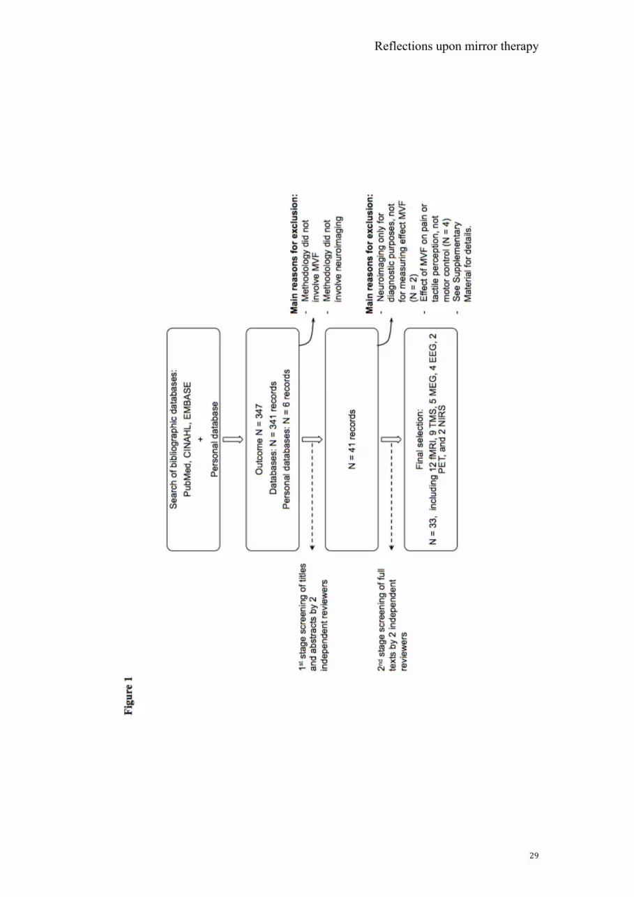
The electronic database search yielded 347 unique articles, of which 33 were
deemed eligible for this systematic review (see Figure 1 for an overview of the
selection process). Across the selected articles, the most commonly used scanning
technique was fMRI (12 studies).37-47 MEG was the neuroimaging modality in 5
article,48-52 EEG in 4,53-56 PET in 2,57,58 and NIRS in 2.59,60 Nine studies
investigated the effect of MVF on cortical activation with TMS47,61-68; 1 study
used both TMS and fMRI.47 In Tables 1 to 4, the included articles and their
methodologies are listed according to modality. The majority of the studies (n =
27) examined immediate modulatory effects when exposed to MVF, of which 22
focused on healthy individuals and 5 on stroke patients (Tables 1-3). In 16 studies,
MVF was provided in a bilateral fashion, that is, not obscuring the active hand.
Six studies assessed neuroplastic changes in response to a bout of practice or an
-Mirror visual feedback could be induced by a real mirror or using a virtual reality environment.!
Reflections upon mirror therapy!
.!
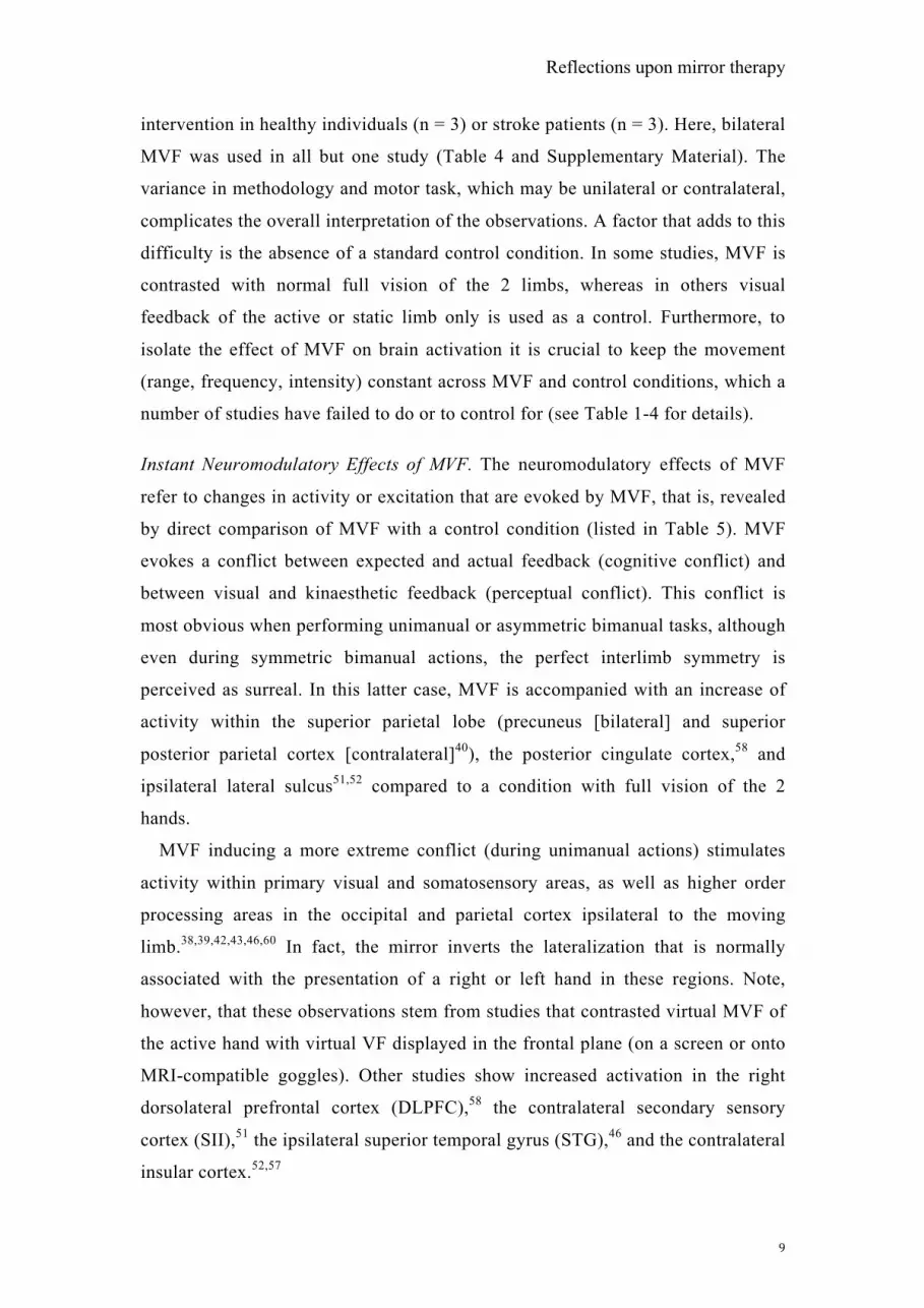
intervention in healthy individuals (n = 3) or stroke patients (n = 3). Here, bilateral
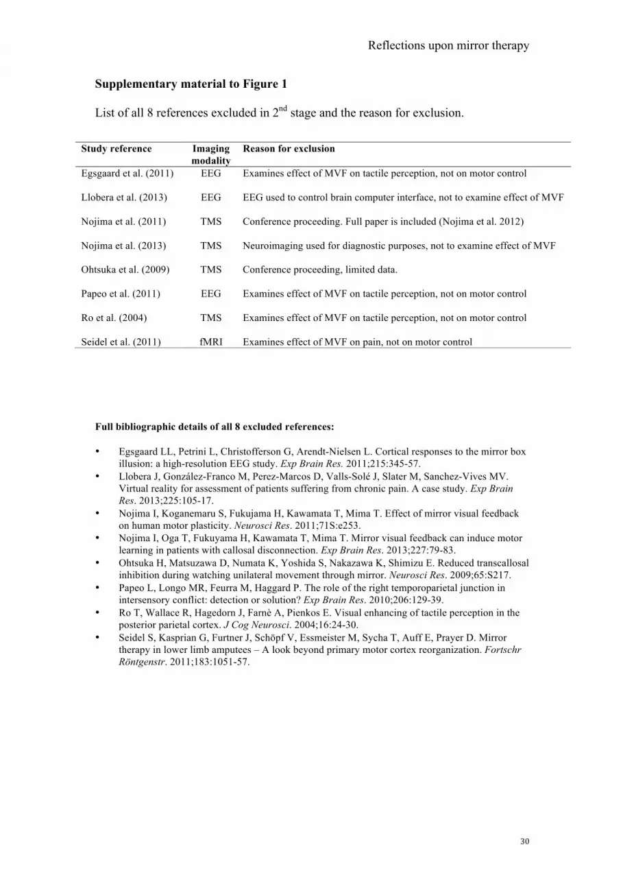
MVF was used in all but one study (Table 4 and Supplementary Material). The
variance in methodology and motor task, which may be unilateral or contralateral,
complicates the overall interpretation of the observations. A factor that adds to this
difficulty is the absence of a standard control condition. In some studies, MVF is
contrasted with normal full vision of the 2 limbs, whereas in others visual
feedback of the active or static limb only is used as a control. Furthermore, to
isolate the effect of MVF on brain activation it is crucial to keep the movement
(range, frequency, intensity) constant across MVF and control conditions, which a
number of studies have failed to do or to control for (see Table 1-4 for details).
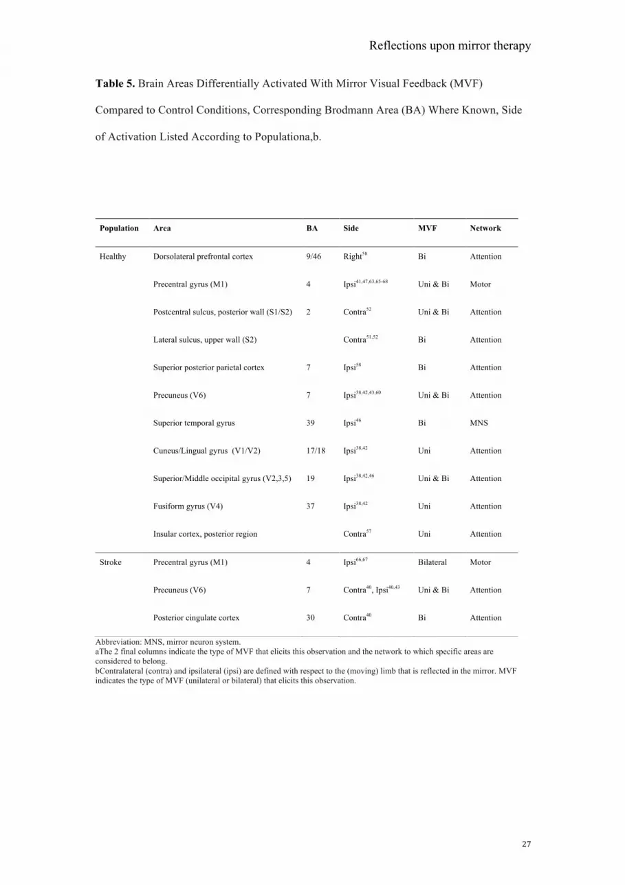
Instant Neuromodulatory Effects of MVF. The neuromodulatory effects of MVF
refer to changes in activity or excitation that are evoked by MVF, that is, revealed
by direct comparison of MVF with a control condition (listed in Table 5). MVF
evokes a conflict between expected and actual feedback (cognitive conflict) and
between visual and kinaesthetic feedback (perceptual conflict). This conflict is
most obvious when performing unimanual or asymmetric bimanual tasks, although
even during symmetric bimanual actions, the perfect interlimb symmetry is
perceived as surreal. In this latter case, MVF is accompanied with an increase of
activity within the superior parietal lobe (precuneus [bilateral] and superior
posterior parietal cortex [contralateral]40), the posterior cingulate cortex,58 and
ipsilateral lateral sulcus51,52 compared to a condition with full vision of the 2
hands.
MVF inducing a more extreme conflict (during unimanual actions) stimulates
activity within primary visual and somatosensory areas, as well as higher order
processing areas in the occipital and parietal cortex ipsilateral to the moving
limb.38,39,42,43,46,60 In fact, the mirror inverts the lateralization that is normally
associated with the presentation of a right or left hand in these regions. Note,
however, that these observations stem from studies that contrasted virtual MVF of
the active hand with virtual VF displayed in the frontal plane (on a screen or onto
MRI-compatible goggles). Other studies show increased activation in the right
dorsolateral prefrontal cortex (DLPFC),58 the contralateral secondary sensory
cortex (SII),51 the ipsilateral superior temporal gyrus (STG),46 and the contralateral
insular cortex.52,57
Reflections upon mirror therapy!
"/!
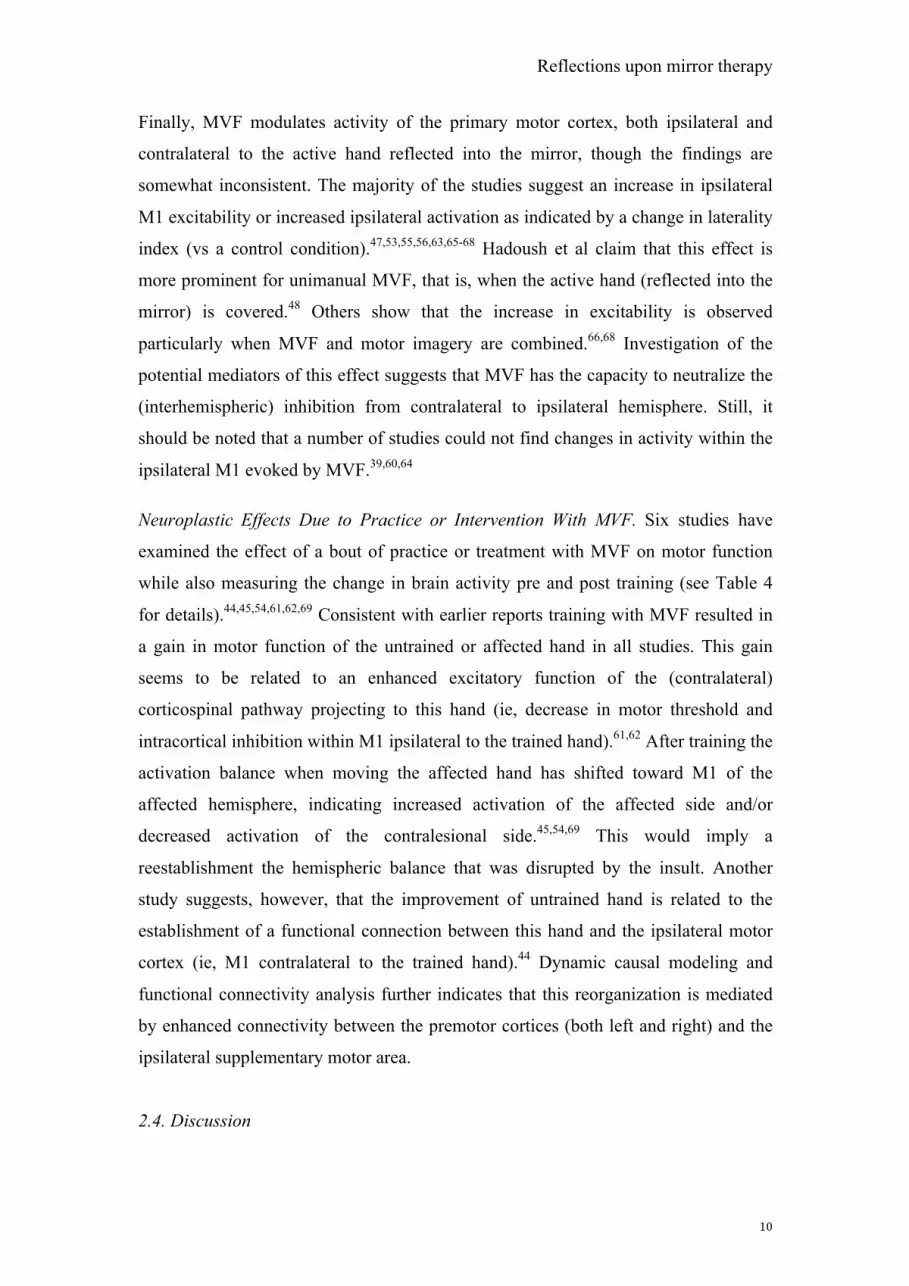
Finally, MVF modulates activity of the primary motor cortex, both ipsilateral and
contralateral to the active hand reflected into the mirror, though the findings are
somewhat inconsistent. The majority of the studies suggest an increase in ipsilateral
M1 excitability or increased ipsilateral activation as indicated by a change in laterality
index (vs a control condition).47,53,55,56,63,65-68 Hadoush et al claim that this effect is
more prominent for unimanual MVF, that is, when the active hand (reflected into the
mirror) is covered.48 Others show that the increase in excitability is observed
particularly when MVF and motor imagery are combined.66,68 Investigation of the
potential mediators of this effect suggests that MVF has the capacity to neutralize the
(interhemispheric) inhibition from contralateral to ipsilateral hemisphere. Still, it
should be noted that a number of studies could not find changes in activity within the
ipsilateral M1 evoked by MVF.39,60,64
Neuroplastic Effects Due to Practice or Intervention With MVF. Six studies have
examined the effect of a bout of practice or treatment with MVF on motor function
while also measuring the change in brain activity pre and post training (see Table 4
for details).44,45,54,61,62,69 Consistent with earlier reports training with MVF resulted in
a gain in motor function of the untrained or affected hand in all studies. This gain
seems to be related to an enhanced excitatory function of the (contralateral)
corticospinal pathway projecting to this hand (ie, decrease in motor threshold and
intracortical inhibition within M1 ipsilateral to the trained hand).61,62 After training the
activation balance when moving the affected hand has shifted toward M1 of the
affected hemisphere, indicating increased activation of the affected side and/or
decreased activation of the contralesional side.45,54,69 This would imply a
reestablishment the hemispheric balance that was disrupted by the insult. Another
study suggests, however, that the improvement of untrained hand is related to the
establishment of a functional connection between this hand and the ipsilateral motor
cortex (ie, M1 contralateral to the trained hand).44 Dynamic causal modeling and
functional connectivity analysis further indicates that this reorganization is mediated
by enhanced connectivity between the premotor cortices (both left and right) and the
ipsilateral supplementary motor area.
2.4. Discussion
Reflections upon mirror therapy!
""!
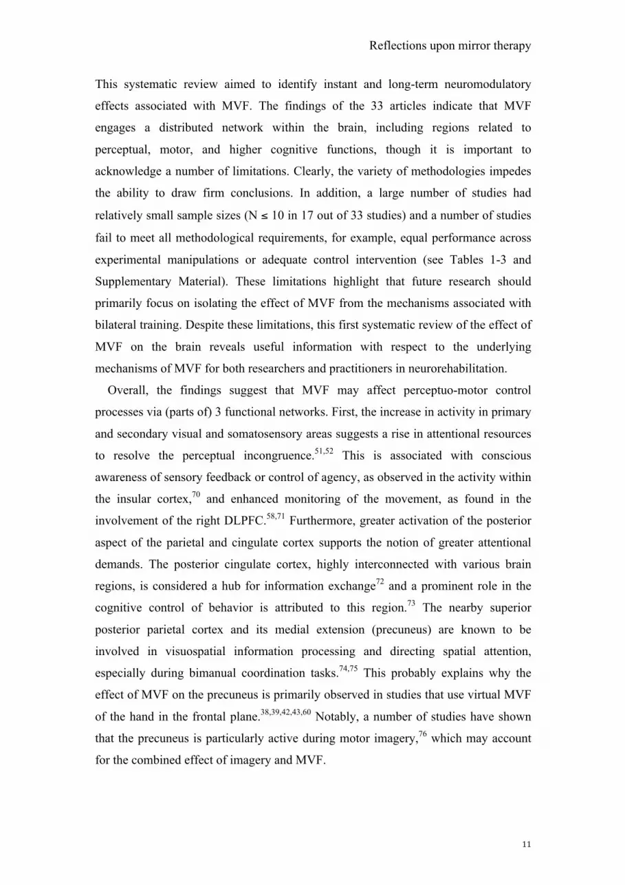
This systematic review aimed to identify instant and long-term neuromodulatory
effects associated with MVF. The findings of the 33 articles indicate that MVF
engages a distributed network within the brain, including regions related to
perceptual, motor, and higher cognitive functions, though it is important to
acknowledge a number of limitations. Clearly, the variety of methodologies impedes
the ability to draw firm conclusions. In addition, a large number of studies had
relatively small sample sizes (N ! 10 in 17 out of 33 studies) and a number of studies
fail to meet all methodological requirements, for example, equal performance across
experimental manipulations or adequate control intervention (see Tables 1-3 and
Supplementary Material). These limitations highlight that future research should
primarily focus on isolating the effect of MVF from the mechanisms associated with
bilateral training. Despite these limitations, this first systematic review of the effect of
MVF on the brain reveals useful information with respect to the underlying
mechanisms of MVF for both researchers and practitioners in neurorehabilitation.
Overall, the findings suggest that MVF may affect perceptuo-motor control
processes via (parts of) 3 functional networks. First, the increase in activity in primary
and secondary visual and somatosensory areas suggests a rise in attentional resources
to resolve the perceptual incongruence.51,52 This is associated with conscious
awareness of sensory feedback or control of agency, as observed in the activity within
the insular cortex,70 and enhanced monitoring of the movement, as found in the
involvement of the right DLPFC.58,71 Furthermore, greater activation of the posterior
aspect of the parietal and cingulate cortex supports the notion of greater attentional
demands. The posterior cingulate cortex, highly interconnected with various brain
regions, is considered a hub for information exchange72 and a prominent role in the
cognitive control of behavior is attributed to this region.73 The nearby superior
posterior parietal cortex and its medial extension (precuneus) are known to be
involved in visuospatial information processing and directing spatial attention,
especially during bimanual coordination tasks.74,75 This probably explains why the
effect of MVF on the precuneus is primarily observed in studies that use virtual MVF
of the hand in the frontal plane.38,39,42,43,60 Notably, a number of studies have shown
that the precuneus is particularly active during motor imagery,76 which may account
for the combined effect of imagery and MVF.
Reflections upon mirror therapy!
"#!
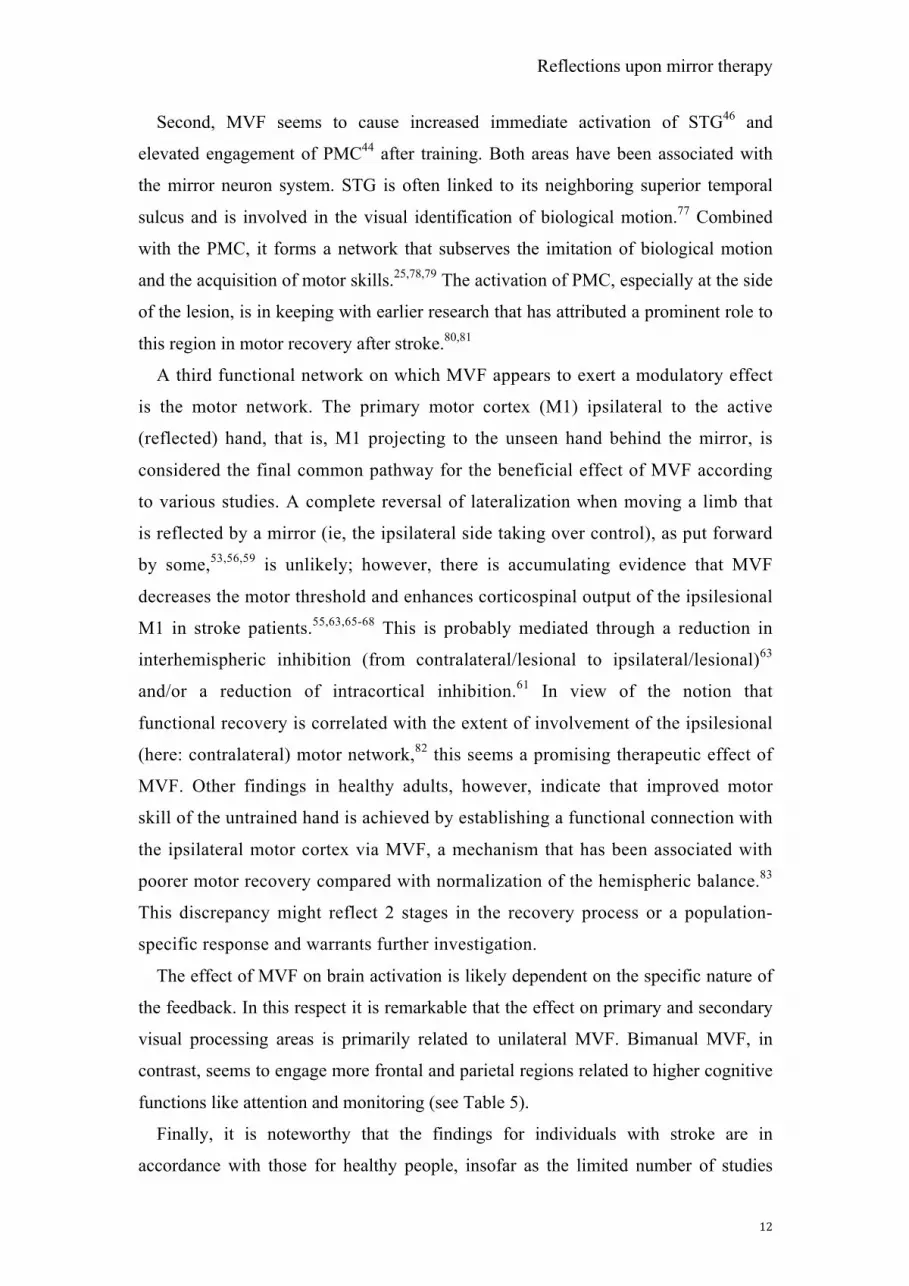
Second, MVF seems to cause increased immediate activation of STG46 and
elevated engagement of PMC44 after training. Both areas have been associated with
the mirror neuron system. STG is often linked to its neighboring superior temporal
sulcus and is involved in the visual identification of biological motion.77 Combined
with the PMC, it forms a network that subserves the imitation of biological motion
and the acquisition of motor skills.25,78,79 The activation of PMC, especially at the side
of the lesion, is in keeping with earlier research that has attributed a prominent role to
this region in motor recovery after stroke.80,81
A third functional network on which MVF appears to exert a modulatory effect
is the motor network. The primary motor cortex (M1) ipsilateral to the active
(reflected) hand, that is, M1 projecting to the unseen hand behind the mirror, is
considered the final common pathway for the beneficial effect of MVF according
to various studies. A complete reversal of lateralization when moving a limb that
is reflected by a mirror (ie, the ipsilateral side taking over control), as put forward
by some,53,56,59 is unlikely; however, there is accumulating evidence that MVF
decreases the motor threshold and enhances corticospinal output of the ipsilesional
M1 in stroke patients.55,63,65-68 This is probably mediated through a reduction in
interhemispheric inhibition (from contralateral/lesional to ipsilateral/lesional)63
and/or a reduction of intracortical inhibition.61 In view of the notion that
functional recovery is correlated with the extent of involvement of the ipsilesional
(here: contralateral) motor network,82 this seems a promising therapeutic effect of
MVF. Other findings in healthy adults, however, indicate that improved motor
skill of the untrained hand is achieved by establishing a functional connection with
the ipsilateral motor cortex via MVF, a mechanism that has been associated with
poorer motor recovery compared with normalization of the hemispheric balance.83
This discrepancy might reflect 2 stages in the recovery process or a population-
specific response and warrants further investigation.
The effect of MVF on brain activation is likely dependent on the specific nature of
the feedback. In this respect it is remarkable that the effect on primary and secondary
visual processing areas is primarily related to unilateral MVF. Bimanual MVF, in
contrast, seems to engage more frontal and parietal regions related to higher cognitive
functions like attention and monitoring (see Table 5).
Finally, it is noteworthy that the findings for individuals with stroke are in
accordance with those for healthy people, insofar as the limited number of studies
Reflections upon mirror therapy!
"$!
allows this comparison (see Table 5). There is evidence of increased activation of
higher order areas involved with attentional processes (precuneus and posterior
cingulate cortex) and the ipsilateral M1.
Part 3: Summary and future directions
Convergent evidence suggests that MVF may be used as a tool to promote
functional recovery in patients with unilateral motor impairments. A systematic
review of neuroimaging research was conducted to test the validity of the 3
hypotheses proposed to explain the positive effects associated with MVF. The
findings of this review, suggesting substantial overlap between MVF-related
activity and regions subserving attention-related processes, confirm that MVF
activates a broad network dedicated to attention and action monitoring (Hypothesis
3). This is consistent with known motor learning principles, which attribute
success of motor practice to attentional focus and cognitive processing.
Furthermore, the positive effect on motor function is associated with facilitation of
M1 contralateral to the affected or untrained hand (here: referred to as ipsilateral
to the moving hand that is mirrored). However, there is also evidence to support a
mechanism that exploits ipsilateral control of the affected limb, which has been
associated with suboptimal recovery after other therapeutic interventions
(Hypothesis 2). Regions that have been linked with the mirror neuron system
(PMC, STG) may play a mediating role in connecting perceptual and motor areas
(Hypothesis 1). Still, the current evidence indicates that MVF therapy is certainly
not a substitute for observational therapy or motor imagery, given that MVF
activates only isolated parts of the MNS. Future research using recent advances in
graph theory may elucidate functional connectivity within and between the
involved networks.84
To date the majority of evidence stems from studies in healthy adult
individuals and the few studies that have examined a patient population only
considered people who survived a stroke. It remains unclear to what extent these
hypotheses may be valid for other clinical conditions for which MVF has been
suggested an adjunct to conventional therapy (eg, SHCP22,85-87 and CRPS14-18).
The finding that MVF may have an impact on multiple functional networks may
Reflections upon mirror therapy!
"%!
mean it can serve as a versatile tool to promote motor recovery, of which the
actual mechanism is dependent on the specific condition or damage. Large-scale
clinical trials that include measurement of brain function and structure are to
examine the efficacy and the underlying mechanisms of MVF in different
populations, and potential differences between them. Although further research is
warranted to fully understand and exploit the potential of MVF in
neurorehabilitation, it is indisputable that MVF can exert a strong modulatory
influence on the motor system.
Reflections upon mirror therapy!
"'!
Acknowledgements
The authors wish to thank Prof. Karen Caeyenberghs and 2 anonymous reviewers for
their advice and suggestions.
Declaration of Conflicting Interest
The author(s) declared no potential conflicts of interest with respect to the research,
authorship, and/or publication of this article.
Funding
The author(s) disclosed receipt of the following financial support for the research,
authorship, and/or publication of this article: This work was partly supported by a
research grant from Sparks, registered charity 1003825 (England & Wales), to Geert
Savelsbergh and Frederik Deconinck (Grant 09MMU01).
Reflections upon mirror therapy!
"*!
References
1. Thompson P, Papadopoulou G, Vassiliou E. The origins of entasis: illusion, aesthetics or engineering? Spat Vis. 2007;20(6):531–43. doi:10.1163/156856807782758359.
2. Gregory RL. Perceptual illusions and brain models. Proc R Soc London Ser B. 1968;171(24):279–96.
3. Ramachandran VS, Rogers-Ramachandran D, Cobb S. Touching the phantom limb. Nature. 1995;377:489–490.
4. Von Holst E, Mittelstaedt H. The Principle of Reafference: Interactions Between the Central Nervous System and the Peripheral Organs. Naturwissenschaften. 1950;37(1950):464–476.
5. Von Helmholtz H. Handbuch der physiologischen Optik. Leipzig: Voss; 1867. 6. Ramachandran VS, Hirstein W. The perception of phantom limbs (The D. O.
Hebb lecture). Brain. 1998;121:1603–1630. 7. Harris AJ. Cortical origin of pathological pain. Lancet. 1999;354(9188):1464–
6. doi:10.1016/S0140-6736(99)05003-5. 8. Gibson JJ. Observations on active touch. Psychol Rev. 1962;69(6):477–91. 9. Mechsner F, Kerzel D, Knoblich G, Prinz W. Perceptual basis of bimanual
coordination. Nature. 2001;414(6859):69–73. doi:10.1038/35102060. 10. Chan BL, Witt R, Charrow AP, et al. Mirror therapy for phantom limb pain. N
pain. Am J Phys Med Rehabil. 2009;88(1):78–81. doi:10.1097/PHM.0b013e318191105b.
12. Ramachandran VS, Altschuler EL. The use of visual feedback, in particular mirror visual feedback, in restoring brain function. Brain. 2009;132(Pt 7):1693–710. doi:10.1093/brain/awp135.
13. Thieme H, Mehrholz J, Pohl M, Behrens J, Dohle C. Mirror therapy for improving motor function after stroke. Cochrane Database Syst Rev. 2012;(3).
14. Selles RW, Schreuders TAR, Stam HJ. Mirror therapy in patients with causalgia (complex regional pain syndrome type II) following peripheral nerve injury: two cases. J Rehabil Med. 2008;40(4):312–4. doi:10.2340/16501977-0158.
15. Karmakar A, Lieberman I. Mirror box therapy for complex regional pain syndrome. Anaesthesia. 2006;61:412–413.
16. Tichelaar YIGV, Geertzen JHB, Keizer D, van Wilgen PC. Mirror box therapy added to cognitive behavioural therapy in three chronic complex regional pain syndrome type I patients: a pilot study. Int J Rehabil Res. 2007;30(2):181–8. doi:10.1097/MRR.0b013e32813a2e4b.
17. Rosen B, Lundborg G. Training with a mirror in rehabilitation of the hand. Scand J Plast Reconstr Surg Hand Surg. 2005;39:104–108.
Reflections upon mirror therapy!
"+!
18. McCabe CS, Haigh RC, Ring EFJ, Halligan PW, Wall PD, Blake DR. A controlled pilot study of the utility of mirror visual feedback in the treatment of complex regional pain syndrome (type 1). Rheumatology. 2002;42:97–101. doi:10.1093/rheumatology/keg041.
19. Sütbeyaz S, Yavuzer G, Sezer N, Koseoglu BF. Mirror therapy enhances lower-extremity motor recovery and motor functioning after stroke: a randomized controlled trial. Arch Phys Med Rehabil. 2007;88(5):555–9. doi:10.1016/j.apmr.2007.02.034.
20. Yavuzer G, Selles R, Sezer N, et al. Mirror therapy improves hand function in subacute stroke: a randomized controlled trial. Arch Phys Med Rehabil. 2008;89(3):393–8. doi:10.1016/j.apmr.2007.08.162.
21. Altschuler EL, Hu J. Mirror therapy in a patient with a fractured wrist and no active wrist extension. Scand J Plast Reconstr Surg Hand Surg. 2008;42(2):110–1. doi:10.1080/02844310701510355.
22. Gygax Jequier M, Schneider P, Newman CJ. Mirror therapy in children with hemiplegia: a pilot study. Dev Med Child Neurol. 2011;53(5):473–6. doi:10.1111/j.1469-8749.2011.03924.x.
23. Brodie EE, Whyte A, Niven CA. Analgesia through the looking-glass? A randomized controlled trial investigating the effect of viewing a “virtual” limb upon phantom limb pain, sensation and movement. Eur J Pain. 2007;11(4):428–36. doi:10.1016/j.ejpain.2006.06.002.
24. Cauraugh JH, Summers JJ. Neural plasticity and bilateral movements: A rehabilitation approach for chronic stroke. Prog Neurobiol. 2005;75(5):309–20. doi:10.1016/j.pneurobio.2005.04.001.
25. Buccino G, Solodkin A, Small SL. Functions of the mirror neuron system: implications for neurorehabilitation. Cogn Behav Neurol. 2006;19(1):55–63.
26. Iacoboni M. Cortical Mechanisms of Human Imitation. Science (80- ). 1999;286(5449):2526–2528. doi:10.1126/science.286.5449.2526.
27. Pomeroy VM, Clark CA, Miller JSG, Baron J-C, Markus HS, Tallis RC. The potential for utilizing the “mirror neurone system” to enhance recovery of the severely affected upper limb early after stroke: a review and hypothesis. Neurorehabil Neural Repair. 2005;19(1):4–13. doi:10.1177/1545968304274351.
28. Stevens J a, Stoykov MEP. Using Motor Imagery in the Rehabilitation of Hemiparesis. Arch Phys Med Rehabil. 2003;84(7):1090–1092. doi:10.1016/S0003-9993(03)00042-X.
29. Grèzes J, Decety J. Functional anatomy of execution, mental simulation, observation, and verb generation of actions: a meta-analysis. Hum Brain Mapp. 2001;12(1):1–19.
30. Ezendam D, Bongers R, Jannink M. Systematic review of the effectiveness of mirror therapy in upper extremity function. Disabil Rehabil. 2009;31(26):2135–2149. doi:10.1080/09638280902887768.
31. Carr LJ, Harrison LM, Stephens JA. Evidence for bilateral innervation of certain homologous motoneurone pools in man. J Physiol. 1994;475(2):217–27.
Reflections upon mirror therapy!
",!
32. Staudt M, Grodd W, Gerloff C, Erb M, Stitz J, Krägeloh-Mann I. Two types of ipsilateral reorganization in congenital hemiparesis: a TMS and fMRI study. Brain. 2002;125(Pt 10):2222–37.
33. Schwerin S, Dewald JPA, Hatzi M, Jovanovich S, Nickeas M, MacKinnon C. Ipsilateral versus contralateral cortical motor projections to a shoulder adductor in chronic hemiparetic stroke: implications for the expression of arm synergies. Exp Brain Res. 2008;185(3):509–519. doi:10.1007/s00221-007-1169-8.Ipsilateral.
34. Benecke R, Meyer BU, Freund HJ. Reorganisation of descending motor pathways in patients after hemispherectomy and severe hemispheric lesions demonstrated by magnetic brain stimulation. Exp Brain Res. 1991;83(2):419–428.
35. Dohle C, Püllen J, Nakaten A, Küst J, Rietz C, Karbe H. Mirror therapy promotes recovery from severe hemiparesis: a randomized controlled trial. Neurorehabil Neural Repair. 2009;23(3):209–17. doi:10.1177/1545968308324786.
36. Taub E, Crago JE, Uswatte G. Constraint-induced movement therapy: A new approach to treatment in physical rehabilitation. Rehabil Psychol. 1998;43(2):152–170. doi:10.1037//0090-5550.43.2.152.
37. Diers M, Christmann C, Koeppe C, Ruf M, Flor H. Mirrored, imagined and executed movements differentially activate sensorimotor cortex in amputees with and without phantom limb pain. Pain. 2010;149(2):296–304. doi:10.1016/j.pain.2010.02.020.
39. Fritzsch C, Wang J, Dos Santos LF, Mauritz K-H, Brunetti M, Dohle C. Different effects of the mirror illusion on motor and somatosensory processing. Restor Neurol Neurosci. 2013.
40. Michielsen ME, Smits M, Ribbers GM, et al. The neuronal correlates of mirror therapy: an fMRI study on mirror induced visual illusions in patients with stroke. J Neurol Neurosurg Psychiatry. 2011;82(4):393–398.
41. Shinoura N, Suzuki Y, Watanabe Y, et al. Mirror therapy activates outside of cerebellum and ipsilateral M1. NeuroRehabilitation. 2008;23(3):245–52.
42. Wang J, Fritzsch C, Bernarding J, et al. A comparison of neural mechanisms in mirror therapy and movement observation therapy. J Rehabil Med. 2013;45:410–3. doi:10.2340/16501977-1127.
43. Wang J, Fritzsch C, Bernarding J, et al. Cerebral activation evoked by the mirror illusion of the hand in stroke patients compared to normal subjects. NeuroRehabilitation. 2013;33(4):593–603.
44. Hamzei F, Läppchen CH, Glauche V, Mader I, Rijntjes M, Weiller C. Functional plasticity induced by mirror training: the mirror as the element connecting both hands to one hemisphere. Neurorehabil Neural Repair. 2012;26(5):484–96. doi:10.1177/1545968311427917.
45. Michielsen ME, Selles RW, van der Geest JN, et al. Motor recovery and cortical reorganization after mirror therapy in chronic stroke patients: a phase II
46. Matthys K, Smits M, JN, et al. Mirror-induced visual illusion of hand movements: a functional magnetic resonance imaging study. Arch Phys Med Rehabil. 2009;90(4):675–681. doi:10.1016/j.apmr.2008.09.571.
47. Merians AS, Tunik E, Fluet GG, Qiu Q, Adamovich S V. Innovative approaches to the rehabilitation of upper extremity hemiparesis using virtual environments. Eur J Phys Rehabil Med. 2009;45(1):123–133.
48. Hadoush H, Mano H, Sunagawa T, Nakanishi K, Ochi M. Optimization of mirror therapy to excite ipsilateral primary motor cortex. NeuroRehabilitation. 2013;32(3):617–624.
49. Tominaga W, Matsubayashi J, Deguchi Y, et al. A mirror reflection of a hand modulates stimulus-induced 20-Hz activity. Neuroimage. 2009;46(2):500–504. doi:10.1016/j.neuroimage.2009.02.021.
50. Tominaga W, Matsubayashi J, Furuya M, et al. Asymmetric activation of the primary motor cortex during observation of a mirror reflection of a hand. PLoS One. 2011;6(11):e28226. doi:10.1371/journal.pone.0028226.
51. Wasaka T, Kakigi R. The effect of unpredicted visual feedback on activation in the secondary somatosensory cortex during movement execution. BMC Neurosci. 2012;13:138. doi:10.1186/1471-2202-13-138.
52. Wasaka T, Kakigi R. Conflict caused by visual feedback modulates activation in somatosensory areas during movement execution. Neuroimage. 2012;59(2):1501–7. doi:10.1016/j.neuroimage.2011.08.024.
53. Touzalin-Chretien P, Ehrler S, Dufour A. Dominance of vision over proprioception on motor programming: evidence from ERP. Cereb Cortex. 2010;20(8):2007–16. doi:10.1093/cercor/bhp271.
54. Bae SH, Jeong WS, Kim KY. Effects of Mirror Therapy on Subacute Stroke Patients’ Brain Waves and Upper Extremity Functions. J Phys Ther Sci. 2012;24(11):1119–1122. doi:10.1589/jpts.24.1119.
55. Praamstra P, Torney L, Rawle CJ, Chris Miall R. Misconceptions about mirror-induced motor cortex activation. Cereb Cortex. 2011;21(8):1935–1940.
56. Touzalin-Chretien P, Dufour A. Motor Cortex Activation Induced by a Mirror!: Evidence From Lateralized Readiness Potentials. J Neurophysiol. 2008;100:19–23. doi:10.1152/jn.90260.2008.
57. Dohle C, Stephan KM, Valvoda JT, et al. Representation of virtual arm movements in precuneus. Exp Brain Res. 2011;208(4):543–555.
58. Fink GR, Marshall JC, Halligan PW, et al. The neural consequences of conflict between intention and the senses. Brain. 1999;122:497–512.
59. Imai I, Takeda K, Shiomi T, Taniguchi T, Kato H. Sensorimotor cortex activation during mirror therapy in healthy right-handed subjects: a study with near-infrared spectroscopy. J Phys Ther Sci. 2008;20(2):141–145.
60. Mehnert J, Brunetti M, Steinbrink J, Niedeggen M, Dohle C. Effect of a mirror-like illusion on activation in the precuneus assessed with functional near-infrared spectroscopy. J Biomed Opt. 2013;18(6):66001.
Reflections upon mirror therapy!
#/!
61. Laeppchen CH, Ringer T, Blessin J, et al. Optical Illusion alters M1 Excitability after Mirror Therapy - a TMS Study. J Neurophysiol. 2012;108(September):2857–2861. doi:10.1152/jn.00321.2012.
62. Nojima I, Mima T, Koganemaru S, Thabit MN, Fukuyama H, Kawamata T. Human motor plasticity induced by mirror visual feedback. J Neurosci. 2012;32(4):1293–300. doi:10.1523/JNEUROSCI.5364-11.2012.
63. Carson RG, Ruddy KL. Vision modulates corticospinal suppression in a functionally specific manner during movement of the opposite limb. J Neurosci. 2012;32(2):646–52. doi:10.1523/JNEUROSCI.4435-11.2012.
64. Funase K, Tabira T, Higashi T, Liang N, Kasai T. Increased corticospinal excitability during direct observation of self-movement and indirect observation with a mirror box. Neurosci Lett. 2007;419(2):108–12. doi:10.1016/j.neulet.2007.04.025.
65. Garry MI, Loftus A, Summers JJ. Mirror, mirror on the wall: viewing a mirror reflection of unilateral hand movements facilitates ipsilateral M1 excitability. Exp Brain Res. 2005;163(1):118–22. doi:10.1007/s00221-005-2226-9.
66. Kang YJ, Ku J, Kim HJ, Park HK. Facilitation of corticospinal excitability according to motor imagery and mirror therapy in healthy subjects and stroke patients. Ann Rehabil Med. 2011;35(6):747–58. doi:10.5535/arm.2011.35.6.747.
67. Kang YJ, Park HK, Kim HJ, et al. Upper extremity rehabilitation of stroke: facilitation of corticospinal excitability using virtual mirror paradigm. J Neuroeng Rehabil. 2012;9:71.
68. Fukumura K, Sugawara K, Tanabe S, Ushiba J, Tomita Y. Influence of mirror therapy on human motor cortex. Int J Neurosci. 2007;117(7):1039–1048.
69. Bhasin A, Padma Srivastava M, Kumaran S, Bhatia R, Mohanty S. Neural interface of mirror therapy in chronic stroke patients: A functional magnetic resonance imaging study. Neurol India. 2013;60:570–576.
70. Farrer C, Frith CD. Experiencing oneself vs another person as being the cause of an action: the neural correlates of the experience of agency. Neuroimage. 2002;15(3):596–603. doi:10.1006/nimg.2001.1009.
71. Mima T, Nagamine T, Nakamura K, Shibasaki H. Attention Modulates Both Primary and Second Somatosensory Cortical Activities in Humans!: A Magnetoencephalographic Study. J Neurophysiol. 1998;80:2215–2221.
72. Hagmann P, Cammoun L, Gigandet X, et al. Mapping the structural core of human cerebral cortex. PLoS Biol. 2008;6(7):e159. doi:10.1371/journal.pbio.0060159.
73. Leech R, Braga R, Sharp DJ. Echoes of the brain within the posterior cingulate cortex. J Neurosci. 2012;32(1):215–22. doi:10.1523/JNEUROSCI.3689-11.2012.
74. Andersen RA. Encoding of intention and spatial location in the posterior parietal cortex. Cereb Cortex. 1995;5(5):457–69.
75. Wenderoth N, Debaere F, Sunaert S, Swinnen SP. The role of anterior cingulate cortex and precuneus in the coordination of motor behaviour. Eur J Neurosci. 2005;22(1):235–46. doi:10.1111/j.1460-9568.2005.04176.x.
Reflections upon mirror therapy!
#"!
76. Cavanna AE, Trimble MR. The precuneus: a review of its functional anatomy and behavioural correlates. Brain. 2006;129(Pt 3):564–83. doi:10.1093/brain/awl004.
77. Schultz J, Imamizu H, Kawato M, Frith CD. Activation of the human superior temporal gyrus during observation of goal attribution by intentional objects. J Cogn Neurosci. 2004;16(10):1695–705. doi:10.1162/0898929042947874.
78. Iacoboni M, Koski LM, Brass M, Bekkering H, Woods RP, Dubeau M. Reafferent copies of imitated actions in the right. Proc Natl Acad Sci USA. 2001;98(24):13995–13999.
79. Schultz J, Friston KJ, O’Doherty J, Wolpert DM, Frith CD. Activation in posterior superior temporal sulcus parallels parameter inducing the percept of animacy. Neuron. 2005;45(4):625–35. doi:10.1016/j.neuron.2004.12.052.
80. Levy CE, Nichols DS, Schmalbrock PM, Keller P, Chakeres DW. Functional MRI evidence of cortical reorganization in upper-limb stroke hemiplegia treated with constraint-induced movement therapy. Am J Phys Med Rehabil. 2001;80(1):4–12.
81. Liepert J, Bauder H, Miltner WHR, Taub E, Weiller C. Treatment-Induced Cortical Reorganization After Stroke in Humans. Stroke. 2000;31(6):1210–1216. doi:10.1161/01.STR.31.6.1210.
82. Calautti C, Baron J-C. Functional neuroimaging studies of motor recovery after stroke in adults: a review. Stroke. 2003;34(6):1553–66. doi:10.1161/01.STR.0000071761.36075.A6.
83. Ward NS, Brown MM, Thompson a J, Frackowiak RSJ. Neural correlates of motor recovery after stroke: a longitudinal fMRI study. Brain. 2003;126(Pt 11):2476–96. doi:10.1093/brain/awg245.
84. Bullmore E, Sporns O. Complex brain networks: graph theoretical analysis of structural and functional systems. Nat Rev Neurosci. 2009;10(3):186–98. doi:10.1038/nrn2575.
85. Feltham MG, Ledebt A, Bennett SJ, Deconinck FJA, Verheul MHG, Savelsbergh GJP. The “mirror box” illusion: effect of visual information on bimanual coordination in children with spastic hemiparetic cerebral palsy. Motor Control. 2010;14(1):68–82.
86. Feltham MG, Ledebt A, Deconinck FJA, Savelsbergh GJP. Mirror visual feedback induces lower neuromuscular activity in children with spastic hemiparetic cerebral palsy. Res Dev Disabil. 2010;31(6):1525–35. doi:10.1016/j.ridd.2010.06.004.
87. Smorenburg ARP, Ledebt A, Deconinck FJA, Savelsbergh GJP. Practicing a matching movement with a mirror in individuals with spastic hemiplegia. Res Dev Disabil. 2013;34:2507–2513.
Reflections upon mirror therapy
! 22
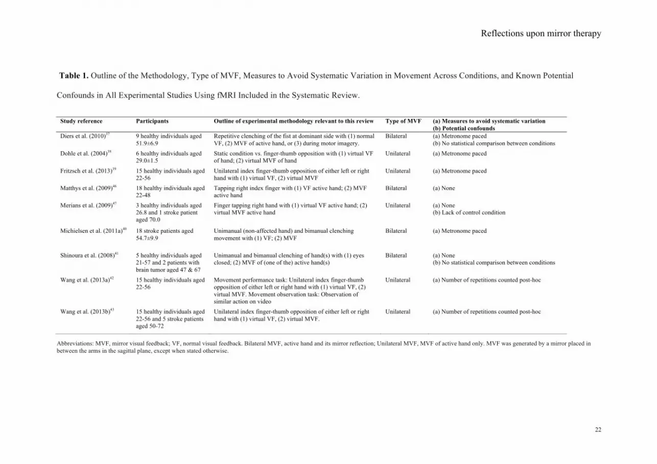
Table 1. Outline of the Methodology, Type of MVF, Measures to Avoid Systematic Variation in Movement Across Conditions, and Known Potential
Confounds in All Experimental Studies Using fMRI Included in the Systematic Review.
Abbreviations: MVF, mirror visual feedback; VF, normal visual feedback. Bilateral MVF, active hand and its mirror reflection; Unilateral MVF, MVF of active hand only. MVF was generated by a mirror placed in between the arms in the sagittal plane, except when stated otherwise.
Study reference Participants Outline of experimental methodology relevant to this review Type of MVF
(a) Measures to avoid systematic variation (b) Potential confounds
Diers et al. (2010)37 9 healthy individuals aged 51.9±6.9
Repetitive clenching of the fist at dominant side with (1) normal VF, (2) MVF of active hand, or (3) during motor imagery.
Bilateral (a) Metronome paced (b) No statistical comparison between conditions
Dohle et al. (2004)38 6 healthy individuals aged 29.0±1.5
Static condition vs. finger-thumb opposition with (1) virtual VF of hand; (2) virtual MVF of hand
Unilateral (a) Metronome paced
Fritzsch et al. (2013)39 15 healthy individuals aged 22-56
Unilateral index finger-thumb opposition of either left or right hand with (1) virtual VF, (2) virtual MVF
Unilateral (a) Metronome paced
Matthys et al. (2009)46 18 healthy individuals aged 22-48
Tapping right index finger with (1) VF active hand; (2) MVF active hand
Bilateral (a) None
Merians et al. (2009)47 3 healthy individuals aged 26.8 and 1 stroke patient aged 70.0
Finger tapping right hand with (1) virtual VF active hand; (2) virtual MVF active hand
Unilateral (a) None (b) Lack of control condition
Michielsen et al. (2011a)40 18 stroke patients aged 54.7±9.9
Unimanual (non-affected hand) and bimanual clenching movement with (1) VF; (2) MVF
Bilateral (a) Metronome paced
Shinoura et al. (2008)41 5 healthy individuals aged 21-57 and 2 patients with brain tumor aged 47 & 67
Unimanual and bimanual clenching of hand(s) with (1) eyes closed; (2) MVF of (one of the) active hand(s)
Bilateral (a) None (b) No statistical comparison between conditions
Wang et al. (2013a)42 15 healthy individuals aged 22-56
Movement performance task: Unilateral index finger-thumb opposition of either left or right hand with (1) virtual VF, (2) virtual MVF. Movement observation task: Observation of similar action on video
Unilateral (a) Number of repetitions counted post-hoc
Wang et al. (2013b)43 15 healthy individuals aged 22-56 and 5 stroke patients aged 50-72
Unilateral index finger-thumb opposition of either left or right hand with (1) virtual VF, (2) virtual MVF.
Unilateral (a) Number of repetitions counted post-hoc
Reflections upon mirror therapy
! 23
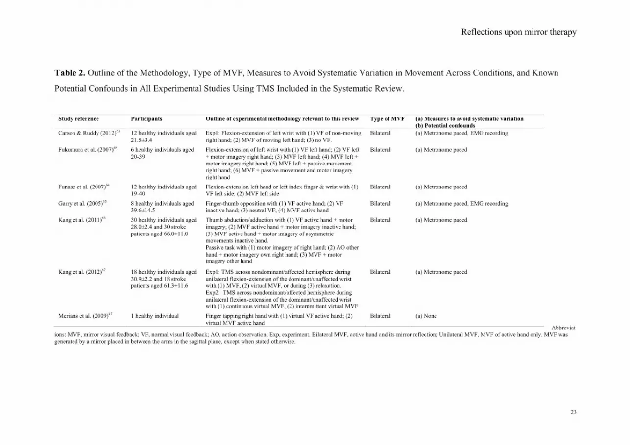
Table 2. Outline of the Methodology, Type of MVF, Measures to Avoid Systematic Variation in Movement Across Conditions, and Known
Potential Confounds in All Experimental Studies Using TMS Included in the Systematic Review.
Abbreviat
ions: MVF, mirror visual feedback; VF, normal visual feedback; AO, action observation; Exp, experiment. Bilateral MVF, active hand and its mirror reflection; Unilateral MVF, MVF of active hand only. MVF was generated by a mirror placed in between the arms in the sagittal plane, except when stated otherwise.
Study reference Participants Outline of experimental methodology relevant to this review Type of MVF
(a) Measures to avoid systematic variation (b) Potential confounds
Exp1: Flexion-extension of left wrist with (1) VF of non-moving right hand; (2) MVF of moving left hand; (3) no VF.
Bilateral (a) Metronome paced, EMG recording
Fukumura et al. (2007)68 6 healthy individuals aged 20-39
Flexion-extension of left wrist with (1) VF left hand; (2) VF left + motor imagery right hand; (3) MVF left hand; (4) MVF left + motor imagery right hand; (5) MVF left + passive movement right hand; (6) MVF + passive movement and motor imagery right hand
Bilateral (a) Metronome paced
Funase et al. (2007)64 12 healthy individuals aged 19-40
Flexion-extension left hand or left index finger & wrist with (1) VF left side; (2) MVF left side
Bilateral (a) Metronome paced
Garry et al. (2005)65 8 healthy individuals aged 39.6±14.5
Finger-thumb opposition with (1) VF active hand; (2) VF inactive hand; (3) neutral VF; (4) MVF active hand
Bilateral (a) Metronome paced, EMG recording
Kang et al. (2011)66 30 healthy individuals aged 28.0±2.4 and 30 stroke patients aged 66.0±11.0
Thumb abduction/adduction with (1) VF active hand + motor imagery; (2) MVF active hand + motor imagery inactive hand; (3) MVF active hand + motor imagery of asymmetric movements inactive hand. Passive task with (1) motor imagery of right hand; (2) AO other hand + motor imagery own right hand; (3) MVF + motor imagery other hand
Bilateral (a) Metronome paced
Kang et al. (2012)67 18 healthy individuals aged 30.9±2.2 and 18 stroke patients aged 61.3±11.6
Exp1: TMS across nondominant/affected hemisphere during unilateral flexion-extension of the dominant/unaffected wrist with (1) MVF, (2) virtual MVF, or during (3) relaxation. Exp2: TMS across nondominant/affected hemisphere during unilateral flexion-extension of the dominant/unaffected wrist with (1) continuous virtual MVF, (2) internmittent virtual MVF
Bilateral (a) Metronome paced
Merians et al. (2009)47 1 healthy individual Finger tapping right hand with (1) virtual VF active hand; (2) virtual MVF active hand
Bilateral (a) None
Reflections upon mirror therapy
! 24
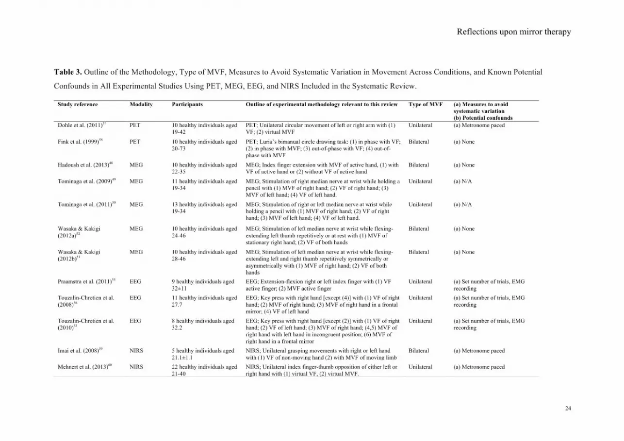
Table 3. Outline of the Methodology, Type of MVF, Measures to Avoid Systematic Variation in Movement Across Conditions, and Known Potential
Confounds in All Experimental Studies Using PET, MEG, EEG, and NIRS Included in the Systematic Review.
Study reference Modality Participants Outline of experimental methodology relevant to this review Type of MVF
(a) Measures to avoid systematic variation (b) Potential confounds
Dohle et al. (2011)57 PET 10 healthy individuals aged 19-42
PET; Unilateral circular movement of left or right arm with (1) VF; (2) virtual MVF
Unilateral (a) Metronome paced
Fink et al. (1999)58 PET 10 healthy individuals aged 20-73
PET; Luria’s bimanual circle drawing task: (1) in phase with VF; (2) in phase with MVF; (3) out-of-phase with VF; (4) out-of-phase with MVF
Bilateral (a) None
Hadoush et al. (2013)48 MEG 10 healthy individuals aged 22-35
MEG; Index finger extension with MVF of active hand, (1) with VF of active hand or (2) without VF of active hand
Bilateral (a) None
Tominaga et al. (2009)49 MEG 11 healthy individuals aged 19-34
MEG; Stimulation of right median nerve at wrist while holding a pencil with (1) MVF of right hand; (2) VF of right hand; (3) MVF of left hand; (4) VF of left hand.
Unilateral (a) N/A
Tominaga et al. (2011)50 MEG 13 healthy individuals aged 19-34
MEG; Stimulation of right or left median nerve at wrist while holding a pencil with (1) MVF of right hand; (2) VF of right hand; (3) MVF of left hand; (4) VF of left hand.
Unilateral (a) N/A
Wasaka & Kakigi (2012a)52
MEG 10 healthy individuals aged 24-46
MEG; Stimulation of left median nerve at wrist while flexing-extending left thumb repetitively or at rest with (1) MVF of stationary right hand; (2) VF of both hands
Bilateral (a) None
Wasaka & Kakigi (2012b)51
MEG 10 healthy individuals aged 28-46
MEG; Stimulation of left median nerve at wrist while flexing-extending left and right thumb repetitively symmetrically or asymmetrically with (1) MVF of right hand; (2) VF of both hands
Bilateral (a) None
Praamstra et al. (2011)55 EEG 9 healthy individuals aged 32±11
EEG; Extension-flexion right or left index finger with (1) VF active finger; (2) MVF active finger
Unilateral (a) Set number of trials, EMG recording
Touzalin-Chretien et al. (2008)56
EEG 11 healthy individuals aged 27.7
EEG; Key press with right hand [except (4)] with (1) VF of right hand; (2) MVF of right hand; (3) MVF of right hand in a frontal mirror; (4) VF of left hand
Unilateral (a) Set number of trials, EMG recording
Touzalin-Chretien et al. (2010)53
EEG 8 healthy individuals aged 32.2
EEG; Key press with right hand [except (2)] with (1) VF of right hand; (2) VF of left hand; (3) MVF of right hand; (4,5) MVF of right hand with left hand in incongruent position; (6) MVF of right hand in a frontal mirror
Unilateral (a) Set number of trials, EMG recording
Imai et al. (2008)59 NIRS 5 healthy individuals aged 21.1±1.1
NIRS; Unilateral grasping movements with right or left hand with (1) VF of non-moving hand (2) with MVF of moving limb
Bilateral (a) Metronome paced
Mehnert et al. (2013)60 NIRS 22 healthy individuals aged 21-40
NIRS; Unilateral index finger-thumb opposition of either left or right hand with (1) virtual VF, (2) virtual MVF.
Unilateral (a) Metronome paced
Reflections upon mirror therapy
! 25
Abbreviations: MVF, mirror visual feedback; VF, normal visual feedback; PET, positron emission topography; MEG, magnetoencephalography; EEG, electroencephalography; NIRS, near infrared spectrometry.
Reflections upon mirror therapy!
"#!
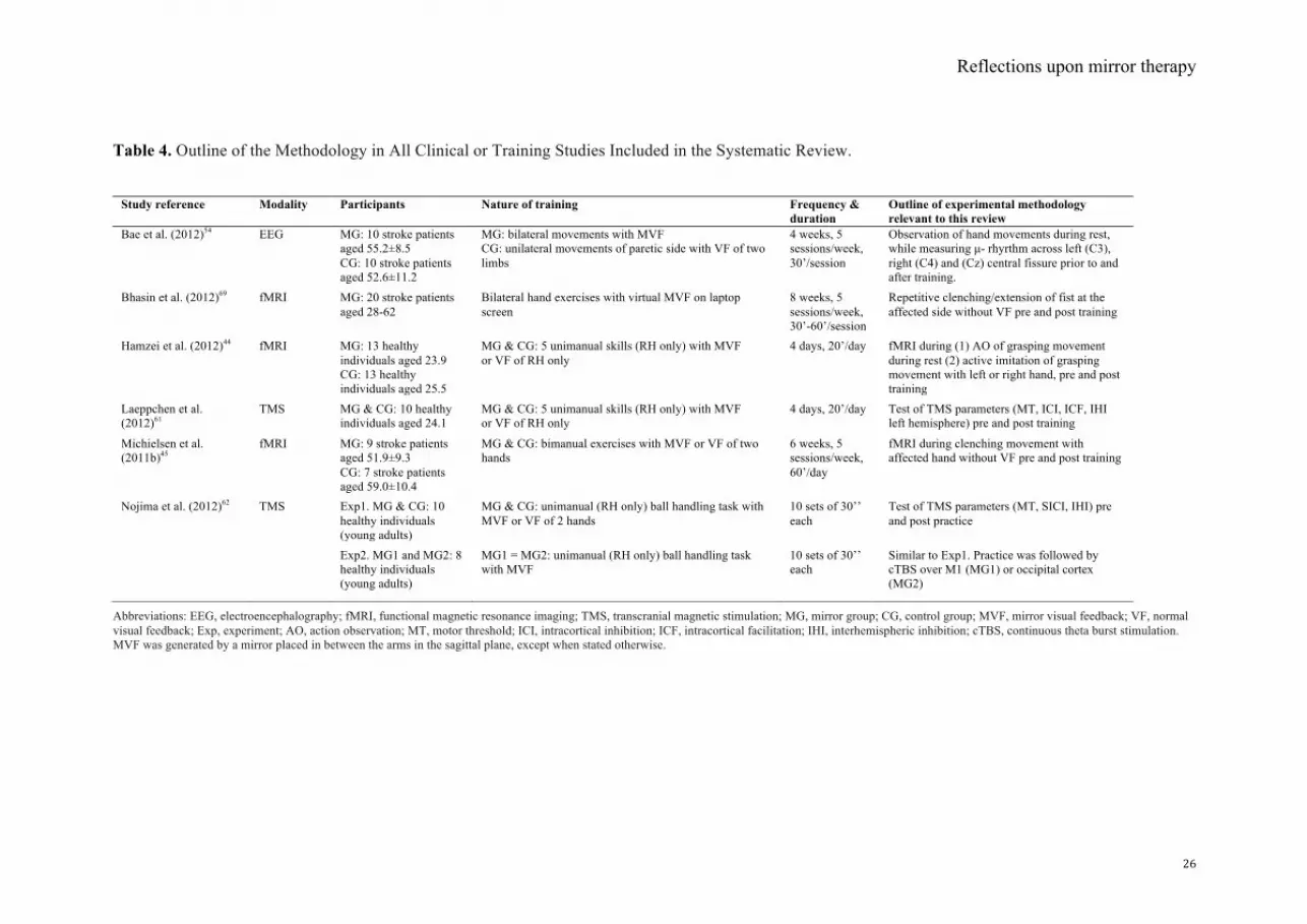
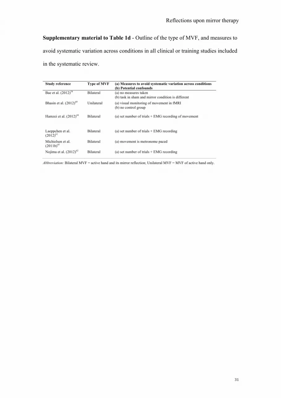
Table 4. Outline of the Methodology in All Clinical or Training Studies Included in the Systematic Review.
Abbreviations: EEG, electroencephalography; fMRI, functional magnetic resonance imaging; TMS, transcranial magnetic stimulation; MG, mirror group; CG, control group; MVF, mirror visual feedback; VF, normal visual feedback; Exp, experiment; AO, action observation; MT, motor threshold; ICI, intracortical inhibition; ICF, intracortical facilitation; IHI, interhemispheric inhibition; cTBS, continuous theta burst stimulation. MVF was generated by a mirror placed in between the arms in the sagittal plane, except when stated otherwise.
Study reference Modality Participants Nature of training Frequency & duration
Outline of experimental methodology relevant to this review
MG: bilateral movements with MVF CG: unilateral movements of paretic side with VF of two limbs
4 weeks, 5 sessions/week, 30’/session
Observation of hand movements during rest, while measuring µ- rhyrthm across left (C3), right (C4) and (Cz) central fissure prior to and after training.
MG & CG: 5 unimanual skills (RH only) with MVF or VF of RH only
4 days, 20’/day fMRI during (1) AO of grasping movement during rest (2) active imitation of grasping movement with left or right hand, pre and post training
Laeppchen et al. (2012)61
TMS MG & CG: 10 healthy individuals aged 24.1
MG & CG: 5 unimanual skills (RH only) with MVF or VF of RH only
4 days, 20’/day Test of TMS parameters (MT, ICI, ICF, IHI left hemisphere) pre and post training
Similar to Exp1. Practice was followed by cTBS over M1 (MG1) or occipital cortex (MG2)
Reflections upon mirror therapy
! 22
Table 1. Outline of the Methodology, Type of MVF, Measures to Avoid Systematic Variation in Movement Across Conditions, and Known Potential
Confounds in All Experimental Studies Using fMRI Included in the Systematic Review.
Abbreviations: MVF, mirror visual feedback; VF, normal visual feedback. Bilateral MVF, active hand and its mirror reflection; Unilateral MVF, MVF of active hand only. MVF was generated by a mirror placed in between the arms in the sagittal plane, except when stated otherwise.
Study reference Participants Outline of experimental methodology relevant to this review Type of MVF
(a) Measures to avoid systematic variation (b) Potential confounds
Diers et al. (2010)37 9 healthy individuals aged 51.9±6.9
Repetitive clenching of the fist at dominant side with (1) normal VF, (2) MVF of active hand, or (3) during motor imagery.
Bilateral (a) Metronome paced (b) No statistical comparison between conditions
Dohle et al. (2004)38 6 healthy individuals aged 29.0±1.5
Static condition vs. finger-thumb opposition with (1) virtual VF of hand; (2) virtual MVF of hand
Unilateral (a) Metronome paced
Fritzsch et al. (2013)39 15 healthy individuals aged 22-56
Unilateral index finger-thumb opposition of either left or right hand with (1) virtual VF, (2) virtual MVF
Unilateral (a) Metronome paced
Matthys et al. (2009)46 18 healthy individuals aged 22-48
Tapping right index finger with (1) VF active hand; (2) MVF active hand
Bilateral (a) None
Merians et al. (2009)47 3 healthy individuals aged 26.8 and 1 stroke patient aged 70.0
Finger tapping right hand with (1) virtual VF active hand; (2) virtual MVF active hand
Unilateral (a) None (b) Lack of control condition
Michielsen et al. (2011a)40 18 stroke patients aged 54.7±9.9
Unimanual (non-affected hand) and bimanual clenching movement with (1) VF; (2) MVF
Bilateral (a) Metronome paced
Shinoura et al. (2008)41 5 healthy individuals aged 21-57 and 2 patients with brain tumor aged 47 & 67
Unimanual and bimanual clenching of hand(s) with (1) eyes closed; (2) MVF of (one of the) active hand(s)
Bilateral (a) None (b) No statistical comparison between conditions
Wang et al. (2013a)42 15 healthy individuals aged 22-56
Movement performance task: Unilateral index finger-thumb opposition of either left or right hand with (1) virtual VF, (2) virtual MVF. Movement observation task: Observation of similar action on video
Unilateral (a) Number of repetitions counted post-hoc
Wang et al. (2013b)43 15 healthy individuals aged 22-56 and 5 stroke patients aged 50-72
Unilateral index finger-thumb opposition of either left or right hand with (1) virtual VF, (2) virtual MVF.
Unilateral (a) Number of repetitions counted post-hoc
Reflections upon mirror therapy
! 23
Table 2. Outline of the Methodology, Type of MVF, Measures to Avoid Systematic Variation in Movement Across Conditions, and Known
Potential Confounds in All Experimental Studies Using TMS Included in the Systematic Review.
Abbreviat
ions: MVF, mirror visual feedback; VF, normal visual feedback; AO, action observation; Exp, experiment. Bilateral MVF, active hand and its mirror reflection; Unilateral MVF, MVF of active hand only. MVF was generated by a mirror placed in between the arms in the sagittal plane, except when stated otherwise.
Study reference Participants Outline of experimental methodology relevant to this review Type of MVF
(a) Measures to avoid systematic variation (b) Potential confounds
Exp1: Flexion-extension of left wrist with (1) VF of non-moving right hand; (2) MVF of moving left hand; (3) no VF.
Bilateral (a) Metronome paced, EMG recording
Fukumura et al. (2007)68 6 healthy individuals aged 20-39
Flexion-extension of left wrist with (1) VF left hand; (2) VF left + motor imagery right hand; (3) MVF left hand; (4) MVF left + motor imagery right hand; (5) MVF left + passive movement right hand; (6) MVF + passive movement and motor imagery right hand
Bilateral (a) Metronome paced
Funase et al. (2007)64 12 healthy individuals aged 19-40
Flexion-extension left hand or left index finger & wrist with (1) VF left side; (2) MVF left side
Bilateral (a) Metronome paced
Garry et al. (2005)65 8 healthy individuals aged 39.6±14.5
Finger-thumb opposition with (1) VF active hand; (2) VF inactive hand; (3) neutral VF; (4) MVF active hand
Bilateral (a) Metronome paced, EMG recording
Kang et al. (2011)66 30 healthy individuals aged 28.0±2.4 and 30 stroke patients aged 66.0±11.0
Thumb abduction/adduction with (1) VF active hand + motor imagery; (2) MVF active hand + motor imagery inactive hand; (3) MVF active hand + motor imagery of asymmetric movements inactive hand. Passive task with (1) motor imagery of right hand; (2) AO other hand + motor imagery own right hand; (3) MVF + motor imagery other hand
Bilateral (a) Metronome paced
Kang et al. (2012)67 18 healthy individuals aged 30.9±2.2 and 18 stroke patients aged 61.3±11.6
Exp1: TMS across nondominant/affected hemisphere during unilateral flexion-extension of the dominant/unaffected wrist with (1) MVF, (2) virtual MVF, or during (3) relaxation. Exp2: TMS across nondominant/affected hemisphere during unilateral flexion-extension of the dominant/unaffected wrist with (1) continuous virtual MVF, (2) internmittent virtual MVF
Bilateral (a) Metronome paced
Merians et al. (2009)47 1 healthy individual Finger tapping right hand with (1) virtual VF active hand; (2) virtual MVF active hand
Bilateral (a) None
Reflections upon mirror therapy
! 24
Table 3. Outline of the Methodology, Type of MVF, Measures to Avoid Systematic Variation in Movement Across Conditions, and Known Potential
Confounds in All Experimental Studies Using PET, MEG, EEG, and NIRS Included in the Systematic Review.
Study reference Modality Participants Outline of experimental methodology relevant to this review Type of MVF
(a) Measures to avoid systematic variation (b) Potential confounds
Dohle et al. (2011)57 PET 10 healthy individuals aged 19-42
PET; Unilateral circular movement of left or right arm with (1) VF; (2) virtual MVF
Unilateral (a) Metronome paced
Fink et al. (1999)58 PET 10 healthy individuals aged 20-73
PET; Luria’s bimanual circle drawing task: (1) in phase with VF; (2) in phase with MVF; (3) out-of-phase with VF; (4) out-of-phase with MVF
Bilateral (a) None
Hadoush et al. (2013)48 MEG 10 healthy individuals aged 22-35
MEG; Index finger extension with MVF of active hand, (1) with VF of active hand or (2) without VF of active hand
Bilateral (a) None
Tominaga et al. (2009)49 MEG 11 healthy individuals aged 19-34
MEG; Stimulation of right median nerve at wrist while holding a pencil with (1) MVF of right hand; (2) VF of right hand; (3) MVF of left hand; (4) VF of left hand.
Unilateral (a) N/A
Tominaga et al. (2011)50 MEG 13 healthy individuals aged 19-34
MEG; Stimulation of right or left median nerve at wrist while holding a pencil with (1) MVF of right hand; (2) VF of right hand; (3) MVF of left hand; (4) VF of left hand.
Unilateral (a) N/A
Wasaka & Kakigi (2012a)52
MEG 10 healthy individuals aged 24-46
MEG; Stimulation of left median nerve at wrist while flexing-extending left thumb repetitively or at rest with (1) MVF of stationary right hand; (2) VF of both hands
Bilateral (a) None
Wasaka & Kakigi (2012b)51
MEG 10 healthy individuals aged 28-46
MEG; Stimulation of left median nerve at wrist while flexing-extending left and right thumb repetitively symmetrically or asymmetrically with (1) MVF of right hand; (2) VF of both hands
Bilateral (a) None
Praamstra et al. (2011)55 EEG 9 healthy individuals aged 32±11
EEG; Extension-flexion right or left index finger with (1) VF active finger; (2) MVF active finger
Unilateral (a) Set number of trials, EMG recording
Touzalin-Chretien et al. (2008)56
EEG 11 healthy individuals aged 27.7
EEG; Key press with right hand [except (4)] with (1) VF of right hand; (2) MVF of right hand; (3) MVF of right hand in a frontal mirror; (4) VF of left hand
Unilateral (a) Set number of trials, EMG recording
Touzalin-Chretien et al. (2010)53
EEG 8 healthy individuals aged 32.2
EEG; Key press with right hand [except (2)] with (1) VF of right hand; (2) VF of left hand; (3) MVF of right hand; (4,5) MVF of right hand with left hand in incongruent position; (6) MVF of right hand in a frontal mirror
Unilateral (a) Set number of trials, EMG recording
Imai et al. (2008)59 NIRS 5 healthy individuals aged 21.1±1.1
NIRS; Unilateral grasping movements with right or left hand with (1) VF of non-moving hand (2) with MVF of moving limb
Bilateral (a) Metronome paced
Mehnert et al. (2013)60 NIRS 22 healthy individuals aged 21-40
NIRS; Unilateral index finger-thumb opposition of either left or right hand with (1) virtual VF, (2) virtual MVF.
Unilateral (a) Metronome paced
Reflections upon mirror therapy
! 25
Abbreviations: MVF, mirror visual feedback; VF, normal visual feedback; PET, positron emission topography; MEG, magnetoencephalography; EEG, electroencephalography; NIRS, near infrared spectrometry.
Reflections upon mirror therapy!
"#!
Table 4. Outline of the Methodology in All Clinical or Training Studies Included in the Systematic Review.
Abbreviations: EEG, electroencephalography; fMRI, functional magnetic resonance imaging; TMS, transcranial magnetic stimulation; MG, mirror group; CG, control group; MVF, mirror visual feedback; VF, normal visual feedback; Exp, experiment; AO, action observation; MT, motor threshold; ICI, intracortical inhibition; ICF, intracortical facilitation; IHI, interhemispheric inhibition; cTBS, continuous theta burst stimulation. MVF was generated by a mirror placed in between the arms in the sagittal plane, except when stated otherwise.
Study reference Modality Participants Nature of training Frequency & duration
Outline of experimental methodology relevant to this review
MG: bilateral movements with MVF CG: unilateral movements of paretic side with VF of two limbs
4 weeks, 5 sessions/week, 30’/session
Observation of hand movements during rest, while measuring µ- rhyrthm across left (C3), right (C4) and (Cz) central fissure prior to and after training.
MG & CG: 5 unimanual skills (RH only) with MVF or VF of RH only
4 days, 20’/day fMRI during (1) AO of grasping movement during rest (2) active imitation of grasping movement with left or right hand, pre and post training
Laeppchen et al. (2012)61
TMS MG & CG: 10 healthy individuals aged 24.1
MG & CG: 5 unimanual skills (RH only) with MVF or VF of RH only
4 days, 20’/day Test of TMS parameters (MT, ICI, ICF, IHI left hemisphere) pre and post training
Similar to Exp1. Practice was followed by cTBS over M1 (MG1) or occipital cortex (MG2)
Reflections upon mirror therapy!
"#!
Table 5. Brain Areas Differentially Activated With Mirror Visual Feedback (MVF)
Compared to Control Conditions, Corresponding Brodmann Area (BA) Where Known, Side
of Activation Listed According to Populationa,b.
!!!
Abbreviation: MNS, mirror neuron system. aThe 2 final columns indicate the type of MVF that elicits this observation and the network to which specific areas are considered to belong. bContralateral (contra) and ipsilateral (ipsi) are defined with respect to the (moving) limb that is reflected in the mirror. MVF indicates the type of MVF (unilateral or bilateral) that elicits this observation.
Figure 1: Flow diagram of the article selection process.
Reflections upon mirror therapy!
"%!
!
Reflections upon mirror therapy!
&'!
Supplementary material to Figure 1
List of all 8 references excluded in 2nd stage and the reason for exclusion.
Full bibliographic details of all 8 excluded references: • Egsgaard LL, Petrini L, Christofferson G, Arendt-Nielsen L. Cortical responses to the mirror box
illusion: a high-resolution EEG study. Exp Brain Res. 2011;215:345-57. • Llobera J, González-Franco M, Perez-Marcos D, Valls-Solé J, Slater M, Sanchez-Vives MV.
Virtual reality for assessment of patients suffering from chronic pain. A case study. Exp Brain Res. 2013;225:105-17.
• Nojima I, Koganemaru S, Fukujama H, Kawamata T, Mima T. Effect of mirror visual feedback on human motor plasticity. Neurosci Res. 2011;71S:e253.
• Nojima I, Oga T, Fukuyama H, Kawamata T, Mima T. Mirror visual feedback can induce motor learning in patients with callosal disconnection. Exp Brain Res. 2013;227:79-83.
• Ohtsuka H, Matsuzawa D, Numata K, Yoshida S, Nakazawa K, Shimizu E. Reduced transcallosal inhibition during watching unilateral movement through mirror. Neurosci Res. 2009;65:S217.
• Papeo L, Longo MR, Feurra M, Haggard P. The role of the right temporoparietal junction in intersensory conflict: detection or solution? Exp Brain Res. 2010;206:129-39.
• Ro T, Wallace R, Hagedorn J, Farnè A, Pienkos E. Visual enhancing of tactile perception in the posterior parietal cortex. J Cog Neurosci. 2004;16:24-30.
• Seidel S, Kasprian G, Furtner J, Schöpf V, Essmeister M, Sycha T, Auff E, Prayer D. Mirror therapy in lower limb amputees – A look beyond primary motor cortex reorganization. Fortschr Röntgenstr. 2011;183:1051-57.
!
!
!
!
Study reference Imaging modality
Reason for exclusion
Egsgaard et al. (2011) EEG Examines effect of MVF on tactile perception, not on motor control Llobera et al. (2013) EEG EEG used to control brain computer interface, not to examine effect of MVF Nojima et al. (2011) TMS Conference proceeding. Full paper is included (Nojima et al. 2012) Nojima et al. (2013) TMS Neuroimaging used for diagnostic purposes, not to examine effect of MVF Ohtsuka et al. (2009) TMS Conference proceeding, limited data. Papeo et al. (2011) EEG Examines effect of MVF on tactile perception, not on motor control Ro et al. (2004) TMS Examines effect of MVF on tactile perception, not on motor control Seidel et al. (2011) fMRI Examines effect of MVF on pain, not on motor control
Reflections upon mirror therapy!
&(!
Supplementary material to Table 1d - Outline of the type of MVF, and measures to
avoid systematic variation across conditions in all clinical or training studies included
in the systematic review.
Abbreviation: Bilateral MVF = active hand and its mirror reflection; Unilateral MVF = MVF of active hand only.
Study reference Type of MVF (a) Measures to avoid systematic variation across conditions (b) Potential confounds
Bae et al. (2012)54 Bilateral (a) no measures taken (b) task in sham and mirror condition is different
Bhasin et al. (2012)69 Unilateral (a) visual monitoring of movement in fMRI (b) no control group
Hamzei et al. (2012)44 Bilateral (a) set number of trials + EMG recording of movement
Laeppchen et al. (2012)61
Bilateral (a) set number of trials + EMG recording
Michielsen et al. (2011b)45
Bilateral (a) movement is metronome paced
Nojima et al. (2012)62 Bilateral (a) set number of trials + EMG recording