1. Select clean, homogeneous, and disease-free seeds (see Note 1).2. Check the viability and homogeneity by performing a germination test (see Note
6).
3. Select seeds with good viability (i.e.>90 %) that allow for the assessment of the
mutagen effects.
4. Separate scions of Jatropha according to the stage of maturity (from bottom to
top: hardwood, semihard, and softwood). This classification is recommended
when cuttings are used to perform the radiosensitivity test (see Note 7).5. Cut scions to ten nodes (meristems) per cutting (see Note 8).6. Tie 10–15 cuttings with a rope into a pack (or put in a transparent plastic bag).
Package volume should not exceed the irradiator chamber.
7. Place plant material in Petri dishes or appropriate containers for irradiation (seeNote 9).
2 Chemical and Physical Mutagenesis in Jatropha curcas 25
2.3.2 In Vitro Material
1. Prepare Petri dishes with two wet Whatman filter papers.
2. Prepare one-node cuttings from freshly micropropagated plantlets (subculture
intervals of 3 weeks).
3. Place cuttings into Petri dishes with wet filter paper (20–30 cuttings per
94 � 16 mm Petri dish).
4. Seal Petri dishes with Parafilm to avoid contamination outside the tissue culture
laboratory.
5. Prepare the regeneration media (MS medium supplemented with growth
regulators).
6. Collect the plant material for mutagenesis (rescued mature embryos, shoot tips
from micropropagated cultures, leaves, meristems, and cotyledons).
2.3.3 Mutagenesis by Chemical Agents
2.3.3.1 EMS Mutagenesis of In Vivo Material (See Note 10)
1. Mix the required volumes of distilled water and 2 % (v/v) DMSO and autoclave
at 120 �C for 15 min. Let the mixture cool to room temperature (RT) (see Note11).
(see Note 4).3. Prepare the concentration series of EMS in water-DMSO mixture (Table 2.1),
including a pure water control treatment.
4. Use a sterile syringe and a 0.2 μm filter to add the required volume of EMS
solution to the sterile water-DMSO mixture (see Note 12). Commence with the
lowest concentration.
5. Shake the EMS-DMSO solution vigorously to form a homogeneous emulsion,
and decant approximately equal volumes into each of bottles (for seeds, seeNote 16) and flat containers (for stem cuttings) labeled with the tissue type,
EMS concentration, and incubation time (see Note 17).
Table 2.1 Example of EMS concentrations chosen for mutagenic treatment (T) of seeds and stem
cuttings of J. curcas (see Note 13)
Plant
material T 1 T 2 T 3 T 4 T 5 T 6
Seeds – – 0.8 %, 1.5 h 1.6 %, 1.5 h 0.8 %, 3 h 1.6 %, 3 h
Stem cuttings 0.4 %, 1.5 h 0.4 %, 3 h 0.8 %, 1.5 h 1.6 %, 1.5 h 0.8 %, 3 h 1.6 %, 3 h
Incubation times were 1.5 h and 3 h (see Note 14). Each treatment should include a control (water-
DMSO) (see Note 15)
26 F. Maghuly et al.
6. Note the time and immerse a separate treatment batch of material into each
solution (0 %, 0.4 %, 0.8 %, and 1.6 %) for the selected incubation time (seeNote 18).
7. After 1.5 h of incubation, quickly but carefully decant each of the 1.5 h
treatment batches, and rinse thoroughly with tap water (at least 3–5 times).
Capture the residual EMS solution and any wash water for disposal in a
hazardous waste container labeled as “Hazardous waste.”
8. Continue incubations of the treatment material selected for 3 h incubation time.
9. After 3 h of incubation, quickly but carefully decant each of the 3 h treatment
batches, and rinse thoroughly with tap water (at least 3–5 times). Capture the
residual EMS solution and any wash water for disposal in a hazardous waste
container labeled as “Hazardous waste.”
10. Detoxify the waste and all unused EMS solution by adding 10 % (w/v) sodium
thiosulfate in a 3:1 ratio by volume. Pour into a designated container (marked
with “Hazardous waste”) and let stand for at least six half-lives (see Note 19).11. Decontaminate the working area with sodium thiosulfate, and dispose
EMS-contaminated items according to laboratory safety rules.
12. Wash plant seeds or stem cuttings in the glasshouse.
13. Mutated seeds and stem cuttings can be immediately planted.
14. Collect data on the germination rate (seed) or sprouting of cuttings and the
survival rate after transplants on a weekly basis.
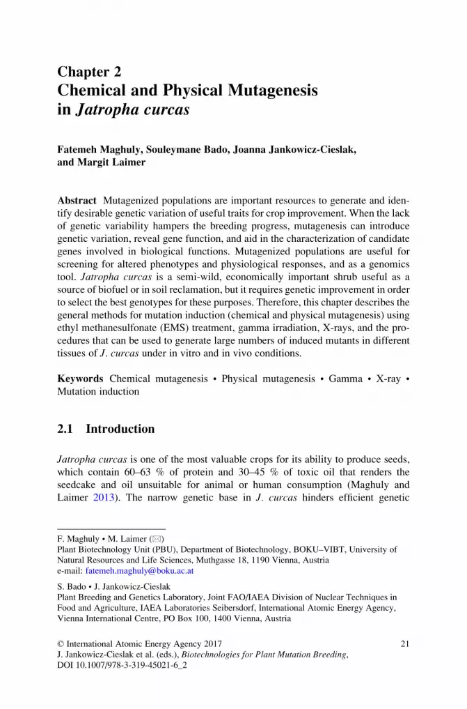
15. Reduce chimerism by producing M2 or higher mutation populations.
16. Screen mutant populations by either phenotypic or genotypic evaluation
(Fig. 2.1.).
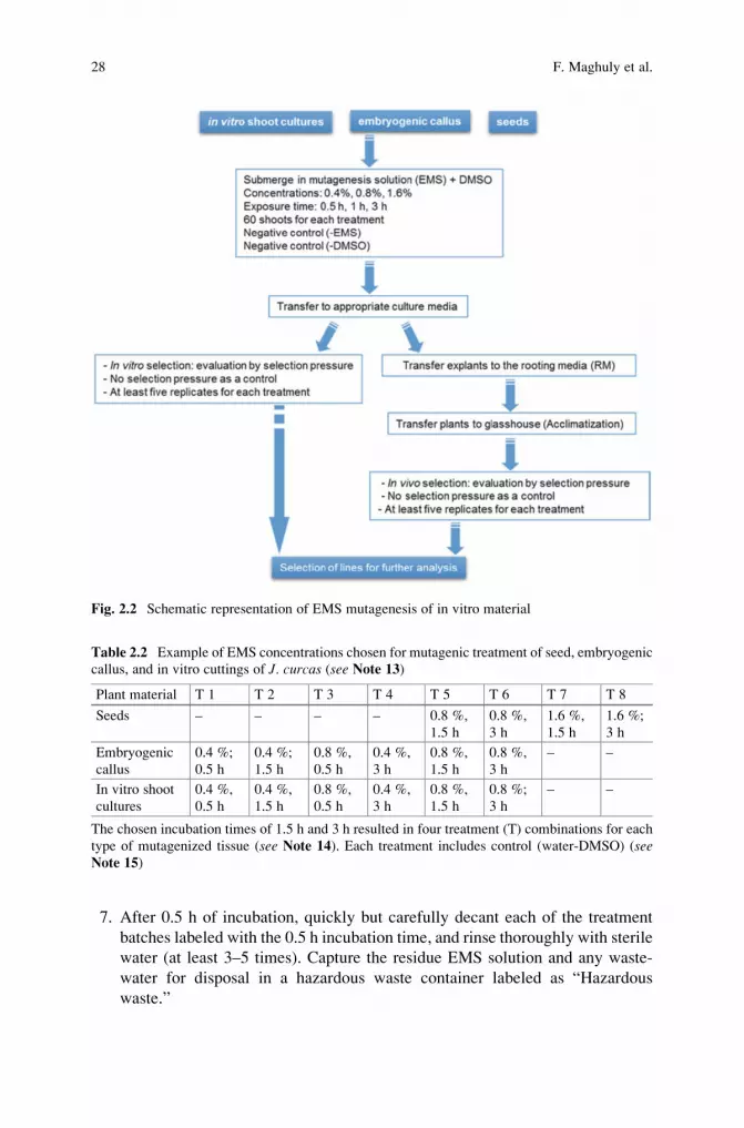
2.3.3.2 EMS Mutagenesis of In Vitro Material (See Notes 10 and 20
and Fig. 2.2)
1. Prepare containers able to accommodate stem cuttings and bottles for seed
mutagenesis. Label them with the incubation time and EMS concentration.
(see Note 4).3. Calculate the final volume of EMS solution needed (Table 2.2, see Note 13).
Mix the required volumes of water and 2 % (v/v) DMSO (see Note 11 and step1 of Sect. 2.3.3.1.).
4. Prepare the concentration series of EMS in water-DMSO mixture, including a
control water treatment (see Note 12, and step 3 of Sect. 2.3.3.1.).
5. Shake the solution vigorously to form a homogeneous emulsion, and decant
approximately equal volumes into each beaker/bottle labeled with the tissue
type, EMS concentration, and incubation time.
6. Note the time. Immerse separate treatment batches of material into each of the
four solutions (0 %, 0.4 %, 0.8 %, and 1.6 %) for three incubation times.
2 Chemical and Physical Mutagenesis in Jatropha curcas 27
7. After 0.5 h of incubation, quickly but carefully decant each of the treatment
batches labeled with the 0.5 h incubation time, and rinse thoroughly with sterile
water (at least 3–5 times). Capture the residue EMS solution and any waste-
water for disposal in a hazardous waste container labeled as “Hazardous
waste.”
Fig. 2.2 Schematic representation of EMS mutagenesis of in vitro material
Table 2.2 Example of EMS concentrations chosen for mutagenic treatment of seed, embryogenic
callus, and in vitro cuttings of J. curcas (see Note 13)
Plant material T 1 T 2 T 3 T 4 T 5 T 6 T 7 T 8
Seeds – – – – 0.8 %,
1.5 h
0.8 %,
3 h
1.6 %,
1.5 h
1.6 %;
3 h
Embryogenic
callus
0.4 %;
0.5 h
0.4 %;
1.5 h
0.8 %,
0.5 h
0.4 %,
3 h
0.8 %,
1.5 h
0.8 %,
3 h
– –
In vitro shoot
cultures
0.4 %,
0.5 h
0.4 %,
1.5 h
0.8 %,
0.5 h
0.4 %,
3 h
0.8 %,
1.5 h
0.8 %;
3 h
– –
The chosen incubation times of 1.5 h and 3 h resulted in four treatment (T) combinations for each
type of mutagenized tissue (see Note 14). Each treatment includes control (water-DMSO) (seeNote 15)
28 F. Maghuly et al.
8. Continue the same way with longer incubation times of 1.5 h and 3 h.
9. Detoxify the waste and unused EMS solution by adding 10 % (w/v) sodium
thiosulfate in a 3:1 ratio by volume. Pour into a designated container (marked
with “Hazardous waste”) and let stand for at least six half-lives (see Note 19
and step 9 of Sect. 2.3.3.1.).
10. Decontaminate working area with sodium thiosulfate, and dispose of EMS
according to laboratory safety rules.
11. After all treatment batches are dispensed, arrange the treated material in each
Petri dish/magenta box containing MS media.
12. Take the cultures to the incubation room with 28 �C and 12 h light.
13. Transfer mutagenized tissue to culture media. After 24 h transfer it again to a
fresh media.
14. Transfer cultures weekly into fresh media to reduce possible accumulation of
phenolic compounds due to the stress of mutagenesis.
15. Thirty days after the treatment, record survival rates of the mutagenized
population.
16. Subculture the growing embryogenic callus and in vitro cutting cultures for
chimera dissolution.
17. Transfer plants to rooting media for acclimatization phase and subsequently to
the glasshouse for further mutant evaluation (Fig. 2.2).
2.3.4 Mutagenesis by Physical Agents
2.3.4.1 Gamma Irradiation of In Vivo Material (See Notes 21–22)
1. Pack seeds in paper envelopes (see Note 23), or tie cuttings with a rope into a
pack (optionally put in a transparent plastic bag) (see Note 7).2. Calculate the exposure time based on the dose rate of gamma cell irradiator that
will be used (Gy/s or Gy/min) (see Note 24).3. Label envelops according to the dose required (Table 2.3) and genotype when
handling more than one genotype. An untreated bag (control) with same
number of seeds will be held in the same conditions as the treated ones.
4. Place the packed seeds in a vacuum desiccator (with 60 % glycerol) for
moisture equilibration.
5. Keep the seeds in vacuum desiccator for 3–7 days. This equilibrates the seed
moisture content to approximately 12–14 %, which is the ideal moisture for
efficient induction of mutation (see Note 25).6. Apply the required dose (Table 2.3) by placing bags into irradiator chamber for
the exposure time to produce the dosage (see Note 7).7. Start gamma cell irradiator (see Note 26).8. Open the lead shielding collar (when gamma cell is at the loading stage).
2 Chemical and Physical Mutagenesis in Jatropha curcas 29
9. Open the sample chamber door.
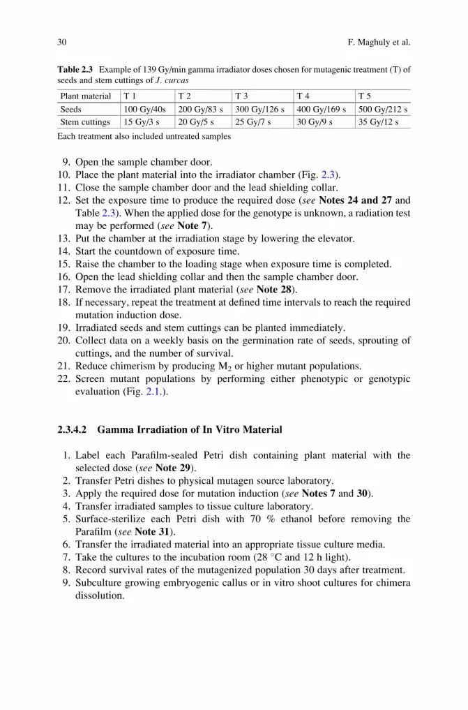
10. Place the plant material into the irradiator chamber (Fig. 2.3).
11. Close the sample chamber door and the lead shielding collar.
12. Set the exposure time to produce the required dose (see Notes 24 and 27 and
Table 2.3). When the applied dose for the genotype is unknown, a radiation test
may be performed (see Note 7).13. Put the chamber at the irradiation stage by lowering the elevator.
14. Start the countdown of exposure time.
15. Raise the chamber to the loading stage when exposure time is completed.
16. Open the lead shielding collar and then the sample chamber door.
17. Remove the irradiated plant material (see Note 28).18. If necessary, repeat the treatment at defined time intervals to reach the required
mutation induction dose.
19. Irradiated seeds and stem cuttings can be planted immediately.
20. Collect data on a weekly basis on the germination rate of seeds, sprouting of
cuttings, and the number of survival.
21. Reduce chimerism by producing M2 or higher mutant populations.
22. Screen mutant populations by performing either phenotypic or genotypic
evaluation (Fig. 2.1.).
2.3.4.2 Gamma Irradiation of In Vitro Material
1. Label each Parafilm-sealed Petri dish containing plant material with the
selected dose (see Note 29).2. Transfer Petri dishes to physical mutagen source laboratory.
3. Apply the required dose for mutation induction (see Notes 7 and 30).
4. Transfer irradiated samples to tissue culture laboratory.
5. Surface-sterilize each Petri dish with 70 % ethanol before removing the
Parafilm (see Note 31).6. Transfer the irradiated material into an appropriate tissue culture media.
7. Take the cultures to the incubation room (28 �C and 12 h light).
8. Record survival rates of the mutagenized population 30 days after treatment.
9. Subculture growing embryogenic callus or in vitro shoot cultures for chimera
dissolution.
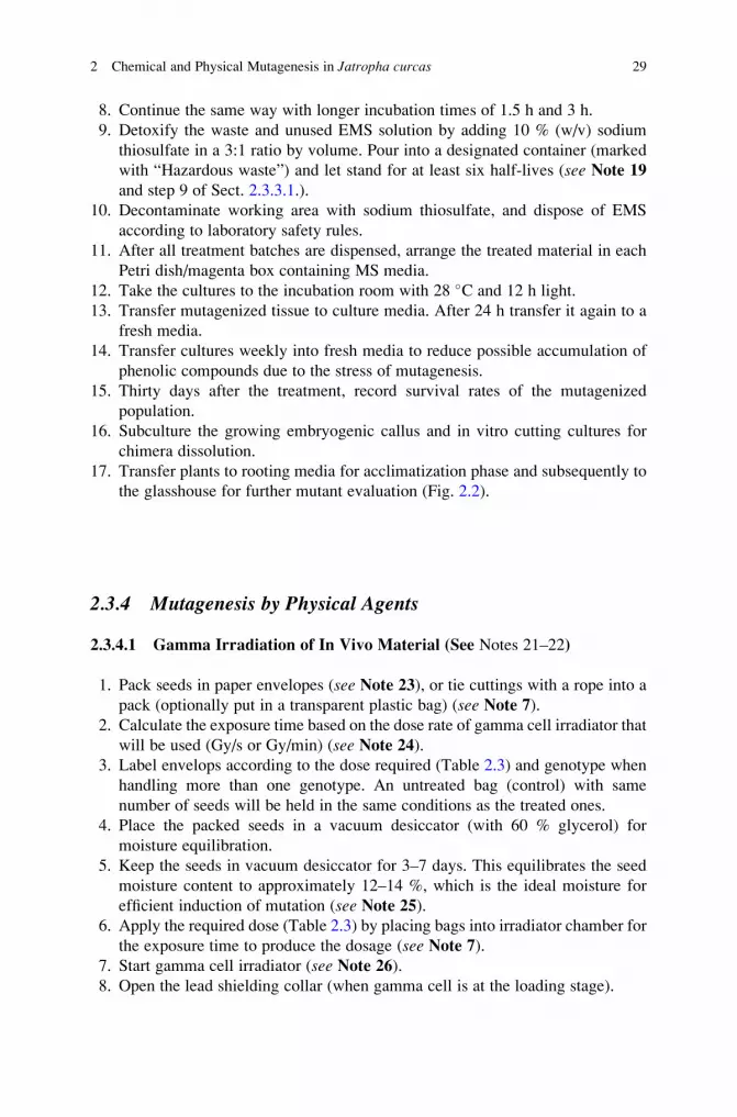
Table 2.3 Example of 139 Gy/min gamma irradiator doses chosen for mutagenic treatment (T) of
seeds and stem cuttings of J. curcas
Plant material T 1 T 2 T 3 T 4 T 5
Seeds 100 Gy/40s 200 Gy/83 s 300 Gy/126 s 400 Gy/169 s 500 Gy/212 s
Stem cuttings 15 Gy/3 s 20 Gy/5 s 25 Gy/7 s 30 Gy/9 s 35 Gy/12 s
Each treatment also included untreated samples
30 F. Maghuly et al.
Fig. 2.3 Process of irradiation using gamma cell irradiator cobalt-60 source
2 Chemical and Physical Mutagenesis in Jatropha curcas 31
10. Transfer plants to rooting media for acclimatization and subsequent transplant
to the glasshouse for further mutant evaluation (Fig. 2.2).
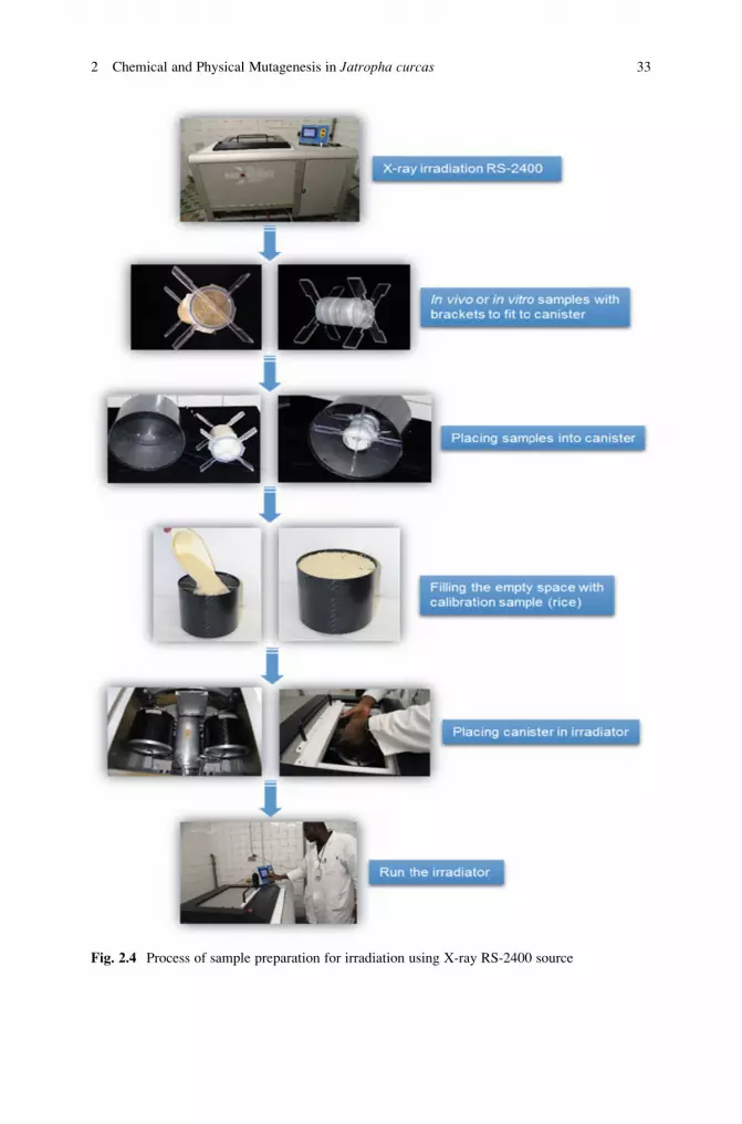
2.3.4.3 X-Rays (See Note 32, Fig. 2.4)
1. Label each Petri dish containing plant material with the selected dose.
2. Place samples into containers and fix them with brackets and move into the
canister.
3. Place canister with adaptors into the center of the irradiator.
4. Create a vacuum by filling the empty space with calibration samples (i.e., rice).
5. Close the canister with the lid.
6. Open the shielding window of the irradiator chamber.
7. Take out the sample platform.
8. Place the canister in one of the five canister holders (make sure that all canisters
are present including the sample canister).
9. Place the platform at the optimal position.
10. Close the shielding window of irradiator chamber.
11. Set the exposure time by putting amount (kW) needed to produce the required
dose (see Note 33).12. Run the irradiator until the monitor displays zero kW.
13. Open the shielding window of the irradiator chamber.
14. Take out the canister containing the irradiated samples.
15. Take off the canister lid.
16. Remove the vacuum samples by dumping the instant rice and do not disturb the
adaptor holding plant material.
17. Remove the samples from the canister and adaptors (see Note 34).18. Take the plant material to appropriate culture media, soil, or storage conditions.
2.4 Further Analyses
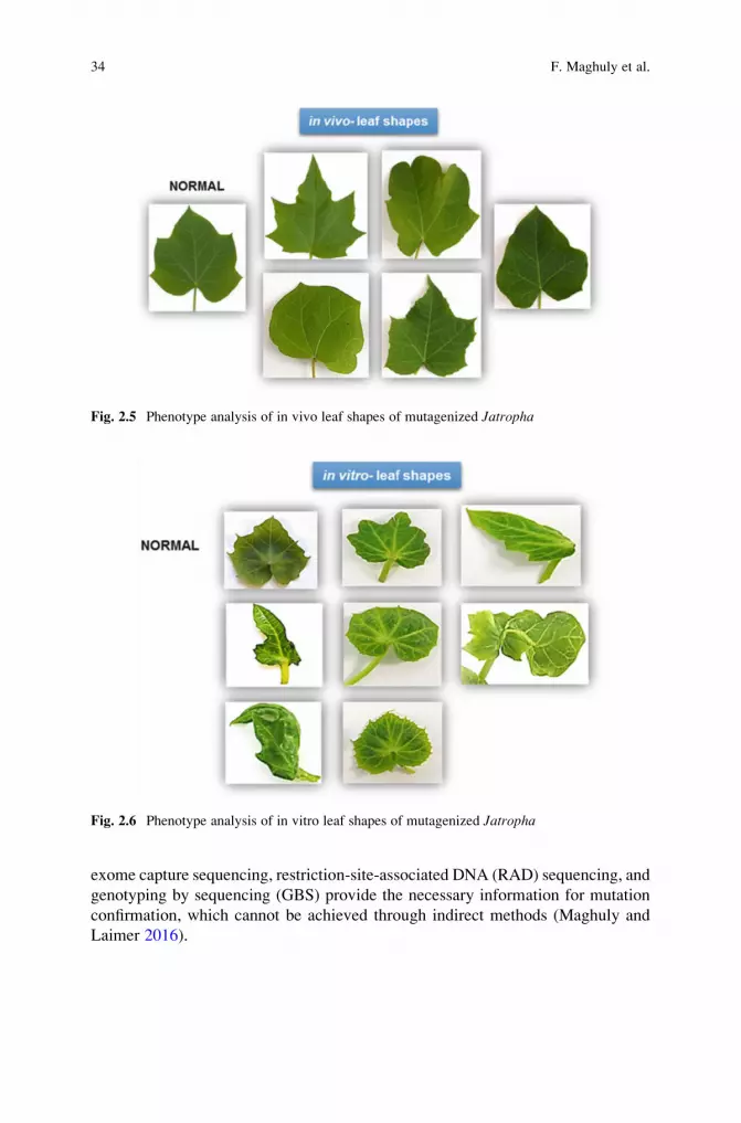
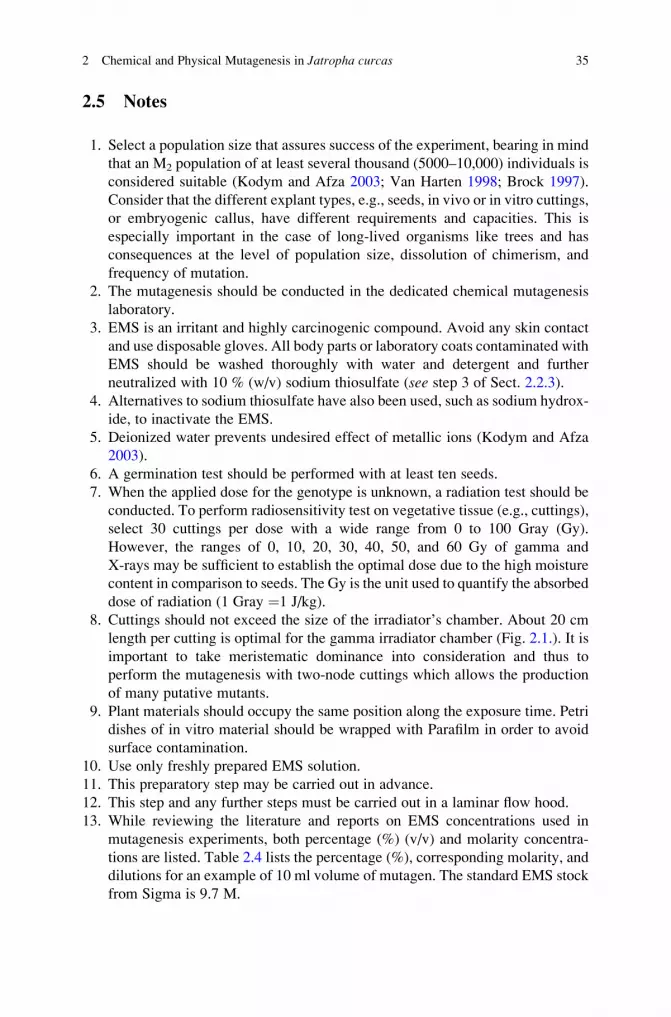
The second generation (and higher) after chimera dissolution of in vivo and in vitro
plants can be screened for the selection of candidate genes based on phenotypes or
genotypes (Figs. 2.5 and 2.6). Mutations can be detected with various direct and
indirect methods such as denaturing high-performance liquid chromatography
(DHPLC), denaturing gradient gel electrophoresis (DGGE), temperature gradient
capillary electrophoresis (TGCE), heteroduplex analysis (HD), the analysis of
single-stranded DNA conformation polymorphism (SSCP), chemical or enzymatic
cleavage of mismatches (CECMs), and Targeting Induced Local Lesions in
Genome (TILLING) (Till et al. 2007). Methods such as whole genome sequencing,
32 F. Maghuly et al.
Fig. 2.4 Process of sample preparation for irradiation using X-ray RS-2400 source
2 Chemical and Physical Mutagenesis in Jatropha curcas 33
exome capture sequencing, restriction-site-associated DNA (RAD) sequencing, and
genotyping by sequencing (GBS) provide the necessary information for mutation
confirmation, which cannot be achieved through indirect methods (Maghuly and
Laimer 2016).
Fig. 2.5 Phenotype analysis of in vivo leaf shapes of mutagenized Jatropha
Fig. 2.6 Phenotype analysis of in vitro leaf shapes of mutagenized Jatropha
34 F. Maghuly et al.
2.5 Notes
1. Select a population size that assures success of the experiment, bearing in mind
that an M2 population of at least several thousand (5000–10,000) individuals is
considered suitable (Kodym and Afza 2003; Van Harten 1998; Brock 1997).
Consider that the different explant types, e.g., seeds, in vivo or in vitro cuttings,
or embryogenic callus, have different requirements and capacities. This is
especially important in the case of long-lived organisms like trees and has
consequences at the level of population size, dissolution of chimerism, and
frequency of mutation.
2. The mutagenesis should be conducted in the dedicated chemical mutagenesis
laboratory.
3. EMS is an irritant and highly carcinogenic compound. Avoid any skin contact
and use disposable gloves. All body parts or laboratory coats contaminated with
EMS should be washed thoroughly with water and detergent and further
neutralized with 10 % (w/v) sodium thiosulfate (see step 3 of Sect. 2.2.3).
4. Alternatives to sodium thiosulfate have also been used, such as sodium hydrox-
ide, to inactivate the EMS.
5. Deionized water prevents undesired effect of metallic ions (Kodym and Afza
2003).
6. A germination test should be performed with at least ten seeds.
7. When the applied dose for the genotype is unknown, a radiation test should be
conducted. To perform radiosensitivity test on vegetative tissue (e.g., cuttings),
select 30 cuttings per dose with a wide range from 0 to 100 Gray (Gy).
However, the ranges of 0, 10, 20, 30, 40, 50, and 60 Gy of gamma and
X-rays may be sufficient to establish the optimal dose due to the high moisture
content in comparison to seeds. The Gy is the unit used to quantify the absorbed
dose of radiation (1 Gray ¼1 J/kg).
8. Cuttings should not exceed the size of the irradiator’s chamber. About 20 cm
length per cutting is optimal for the gamma irradiator chamber (Fig. 2.1.). It is
important to take meristematic dominance into consideration and thus to
perform the mutagenesis with two-node cuttings which allows the production
of many putative mutants.
9. Plant materials should occupy the same position along the exposure time. Petri
dishes of in vitro material should be wrapped with Parafilm in order to avoid
surface contamination.
10. Use only freshly prepared EMS solution.
11. This preparatory step may be carried out in advance.
12. This step and any further steps must be carried out in a laminar flow hood.
13. While reviewing the literature and reports on EMS concentrations used in
mutagenesis experiments, both percentage (%) (v/v) and molarity concentra-
tions are listed. Table 2.4 lists the percentage (%), corresponding molarity, and
dilutions for an example of 10 ml volume of mutagen. The standard EMS stock
from Sigma is 9.7 M.
2 Chemical and Physical Mutagenesis in Jatropha curcas 35
14. Estimate the time needed to conduct the entire experiment with various tissue
types. In case you are not confident in doing the mutagenesis on all tissue types,
split the plant material into batches for separate days treating one tissue type
per day.
15. Some protocols call for the addition of DMSO, which may make the mutagen
more miscible.
16. Presoaking seeds increases infusion of the mutagen into the embryo. The time
of presoaking depends on the seed coat.
17. The amount of mutagen solution should be enough to cover the whole tissue, to
provide a constant concentration of mutagen throughout the treatment, and to
expose all samples similarly.
18. If duration of treatment is longer than half-life of the EMS (see Note 19), thesolution should be replaced with a freshly prepared mutagen solution.
19. The half-life of EMS in water at pH 7 at 20 �C is 93 h and at 30 �C is 26 h
(Kodym and Afza 2003). For EMS in a 10 % sodium thiosulfate solution, the
half-life is 1.4 h at 20 �C and 1 h at 25 �C.20. Somatic embryogenesis is an excellent system for plant propagation and
mutation induction because somatic embryos originate from single cells.
Somatic embryos can be produced on a large scale in cell suspension cultures
in Erlenmeyer flasks or in bioreactors. Somatic embryogenesis limits chimeras
among in vitro plantlets making embryogenic callus an ideal plant material for
mutation induction. Somatic embryos of Jatropha can be induced by direct
somatic embryogenesis or indirect somatic embryogenesis (Kalimuthu
et al. 2007). The difference between the two strategies is whether plants are
regenerated directly from embryos or from an intervening callus phase.
21. Gamma ray mutagenesis may be performed using different facilities, such as
gamma cell irradiator, gamma phytotron, gamma house, or gamma field. The
gamma cell irradiator with cobalt-60 or cesium-137 as radioactive source is the
most commonly available equipment worldwide.
22. Radioactivity is mutagenic and carcinogenic. It should be handled by trained
personnel in a suitable laboratory. The safety precautions for exposing plant
material to a gamma irradiation source must be strictly observed.
23. Perform radiosensitivity test for minimum of ten seeds per bag with at least
three replications per dose, or 100 seeds per bag and per dose.
Table 2.4 Examples of the concentration EMS in molarity or percentage in 10 ml final volume
Molarity (mM) % Amount to add to a final volume of 10 ml (in microliters)
9.7 0.10 10
19.4 0.20 20
22.5 0.23 23
25 0.26 25.7
27.5 0.28 28.3
29 0.30 30
48.5 0.50 50
36 F. Maghuly et al.
24. Exposure time is equal to the required dose divided by the dose rate of the day.
25. The amount of water has an important impact on the irradiation efficiency. A
decreased amount of seed moisture content will increase the mutation fre-
quency. Therefore, it is necessary to equilibrate the seed moisture content
before the irradiation process.
26. Take care to observe all safety precautions before exposing tissues to
irradiation.
27. For each exposure, dosimetry was performed following the Gafchromic dosim-
etry system. One 20 by 20 mm (for X-ray) film dosimeter is placed in a small
paper envelope labeled with requested dose and then placed in the center of
samples before each exposure. The optical density measurement is performed
on a radiochromic reader 24 h after irradiation, and the dose was calculated
according to the calibration Gafchromic dosimetry film with their optical
density. That allows the estimation of the absorbed dose by the sample exposed
to X-ray.
28. The irradiated samples are safe to be held in hands because the sample chamber
isolates the plant material from the source and there is no surface
contamination.
29. A Petri dish with untreated cuttings (control) has to be prepared and kept in the
same conditions as the treated samples.
30. Incubate somatic embryos at the appropriate conditions according to direct or
indirect techniques.
31. Observe general rules for plant tissue culture practices.
Acknowledgments Authors wish to thank FWF, FFG, and Bioplant R&D for the financial
support. Irradiation and chemical mutagenesis experiments described in this protocol were carried
out at the IAEA Laboratories, Seibersdorf, Austria.
Open Access This chapter is distributed under the terms of the Creative Commons Attribution-
Noncommercial 2.5 License (http://creativecommons.org/licenses/by-nc/2.5/) which permits any
noncommercial use, distribution, and reproduction in any medium, provided the original author
(s) and source are credited.
The images or other third party material in this chapter are included in the work’s Creative
Commons license, unless indicated otherwise in the credit line; if such material is not included in
the work’s Creative Commons license and the respective action is not permitted by statutory
regulation, users will need to obtain permission from the license holder to duplicate, adapt or
reproduce the material.
References
Bado S, Kozak K, Sekander H, Alhajaj N, Ghanim A, Forster BP, Laimer M (2013) Resurgence of
X-rays in mutation breeding. Plant Genetics and Breeding Technologies; Plant Diseases and
Resistance Mechanisms: Proceedings, February 18–20, Vienna, Austria. Medimond—
2 Chemical and Physical Mutagenesis in Jatropha curcas 37
Monduzzi Editore international Proceedings Division, Pianoro, Italy, p. 13–16. ISBN 978–88–
7587-682-1.
Bado S, Forster BP, Nielen S, Ghanim A, Lagoda PJL, Till BJ, Laimer M (2015) Plant mutation
breeding: current progress and future assessment. In: Janick J (ed) Plant Breeding Reviews, vol
39. Wiley, New Jersey
Brock RD (1997) When to use mutations in plant breeding. In: Manual on mutation breeding, 2nd
edn. IAEA technical reports series 119, 213–219
Carels N (2013) Towards the domestication of Jatropha: the integration of science. In: Bahadur B,
Sujatha M, Carels N (eds) Jatropha, challenges for a new energy crop, vol 2. Springer,
New York, pp. 263–299
Esnault MA, Legue F, Chenal C (2010) Ionizing radiation: advances in plant response. Environ
Exp Bot 68:231–237
Greene EA, Codomo CA, Taylor NE, Henikoff JG, Till BJ, McCallum CM, Comai L, Henikoff S
(2003) Spectrum of chemically induced mutations from a large-scale reverse-genetic screen in
Arabidopsis. Genetics 164:731–740
Kalimuthu K, Paulsamy S, Senthilkumar R, Sathya M (2007) In vitro propagation of the biodiesel
plant Jatropha curcas L. Plant Tissue Cult & Biotech 17:137–147
Kim, Y., Schumaker, K. S., Zhu, J. K. (2006) EMS mutagenesis of Arabidopsis. In: J. Salinas and
J. J. Sanchez-Serrano (Eds) Methods in molecular biology, Arabidopsis protocols, Second
Edition, vol. 323, Humana Press Inc., Totowa, NJ.
Kodym A, Afza R (2003) Physical and chemical mutagenesis. Methods Mol Biol 236:189–204
Krieg DR (1963) Ethyl methanesulfonate-induced reversion of bacteriophage T4rII mutants.
Genetics 48:561–580
Lagoda PJL (2012) Effects of radiation on living cells and plants. In: Shu QY, Forster BF,
Nakagawa H (eds) Plant mutation breeding and biotechnology. CABI/FAO, Oxforedshsire,
UK/Rome, pp. 123–134
Maghuly F, Laimer M (2013) Jatropha curcas, a biofuel crop: functional genomics for under-
standing metabolic pathways and genetic improvement. J Biotechnol 8:1172–1182
Maghuly F, Laimer M (2016) Forward and reverse genetics for the improvement of Jatrophacurcas. Springer-Verlag.
Mba C, Afza R, Bado S, Jain SM (2010) Induced mutagenesis in plants using physical and
chemical agents. In: Davey MR, Anthony P (eds) Plant cell culture: essential methods.
Wiley, Chichester, UK, pp. 111–130
Mba C, Afza R, Shu QY (2012) Mutagenic radiations: X-rays, ionizing particles and ultraviolet.
In: Shu QY, Forster BF, Nakagawa H (eds) Plant mutation breeding and biotechnology. CAB
Direct, 83–106. 10.1079/9781780640853.0083.McCallum CM, Comai L, Greene EA, Henikoff S (2000) Targeted screening for induced muta-
tions. Nat Biotech 18:455–457
Morita R, Kusaba M, Iida S, Yamaguchi H, Nishio T, Nishimura M (2009) Molecular character-
ization of mutations induced by gamma irradiation in rice. Genes Genet Sys 84:361–370
Pathirana R (2011) Plant mutation breeding in agriculture. CAB Reviews: perspectives in agri-
culture, veterinary science, nutrition and natural resources 6:1–20
Till BJ, Cooper J, Tai TH, Colowit P, Greene EA, Henikoff S, Comai L (2007) Discovery of
chemically induced mutations in rice by TILLING. BMC Plant Biol 7:19–30
Van Harten AM (1998) Mutation breeding theory and practical applications. Cambrige University