Intra-ampullary Papillary-Tubular Neoplasm (IAPN):Characterization of Tumoral Intraepithelial Neoplasia OccurringWithin the Ampulla:A Clinicopathologic Analysis of 82 Cases
Nobuyuki Ohike, MD, PhD*,†, Grace E. Kim, MD‡, Takuma Tajiri, MD, PhD§, AlyssaKrasinskas, MD∥, Olca Basturk, MD¶, Ipek Coban, MD*, Sudeshna Bandyopadhyay, MD#,Toshio Morohoshi, MD, PhD†, Michael Goodman, MD, MPH**, David A. Kooby, MD††, JuanM. Sarmiento, MD††, and N. Volkan Adsay, MD*
*Department of Pathology††Department of Surgery, Emory University Hospital**Department of Epidemiology, Emory University, Rollins School of Public Health, Atlanta, GA‡Department of Pathology, University of California San Francisco, San Francisco, CA∥Department of Pathology, University of Pittsburgh, PA¶Department of Pathology, Memorial Sloan-Kettering Cancer Center, NY#Department of Pathology, The Karmanos Cancer Institute and Wayne State University, MI†First Department of Pathology, Showa University, Tokyo§Department of Pathology, Showa University, Fujigaoka Hospital, Yokohama, Japan
AbstractBackground—There has been no uniform terminology for systematic analysis of mass-formingpreinvasive neoplasms (which we term tumoral intraepithelial neoplasia) that occur specificallywithin the ampulla. Here, we provide a detailed analysis of these neoplasms, which we propose torefer to as intra-ampullary papillary-tubular neoplasm (IAPN).
Materials and Methods—Three hundred and seventeen glandular neoplasms involving theampulla were identified through a review of 1469 pancreatoduodenectomies and 11ampullectomies. Eighty-two neoplasms characterized by substantial preinvasive exophyticcomponent that grew almost exclusively (>75%) within the ampulla (in the ampullary channel orintraampullary portions of the very distal segments of the common bile duct or pancreatic duct)were analyzed.
Results—(1) Clinical: The mean age was 64 years, male/female ratio was 2.4, and mean tumorsize was 2.7 cm. (2) Pathology: The tumors had a mixture of both papillary and tubular growth(each constituting at least 25% of the lesion) in 57%; predominantly (>75%) papillary in 23%, andpredominantly (>75%) tubular in 20%. High-grade dysplasia was present in 94% of cases, ofwhich 39% showed focal (<25% of the lesion), 28% showed substantial (25% to 75%), and 27%
NIH Public AccessAuthor ManuscriptAm J Surg Pathol. Author manuscript; available in PMC 2011 September 7.
Published in final edited form as:Am J Surg Pathol. 2010 December ; 34(12): 1731–1748. doi:10.1097/PAS.0b013e3181f8ff05.
NIH
-PA Author Manuscript
NIH
-PA Author Manuscript
NIH
-PA Author Manuscript
showed extensive (>75%) high-grade dysplasia. In terms of cell-lineage morphology, 45% had amixture of patterns. However, when evaluated with a forced-binary approach as intestinal (INT)versus gastric/pancreatobiliary (GPB) based on the predominant pattern, 74% were classified asINT and 26% as GPB. (3) Immunohistochemistry: Percent sensitivity/specificity of cell-lineagemarkers were, for INT phenotype: MUC2 85/78 and CDX2 94/61; and for GBP: MUC1 89/79,MUC5AC 95/69, and MUC6 83/76, respectively. Cytokeratin 7 and 20 were coexpressed in morethan half. (4) Invasive carcinoma: In 64 cases (78%), there was an associated invasive carcinoma.Size of the tumor and amount of dysplasia correlated with the incidence of invasion. Invasivecarcinoma was of INT-type in 58% and of pancreatobiliary-type in 42%. Cell lineage in theinvasive component was the same as that of the preinvasive component in 84%. All discrepantcases were pancreatobiliary-type invasions, which occurred in INT-type preinvasive lesions. (5)Outcome: The overall survival of invasive cases were significantly worse than that of noninvasiveones (57% vs. 93%; P = 0.01); and 3 years, 69% versus 100% (P = 0.08); and 5 years, 45% versus100% (P = 0.07), respectively. When compared with 166 conventional invasive carcinomas of theampullary region, invasive IAPNs had significantly better prognosis with a mean survival of 51versus 31 months (P<0.001) and the 3-year survival of 69% versus 44% (P<0.01).
Conclusions—Tumoral intraepithelial neoplasia occurring within the ampulla are highlyanalogous to pancreatic or biliary intraductal papillary and tubular neoplasms as evidenced bytheir papillary and/or tubular growth, variable cell lineage, and spectrum of dysplastic change(adenoma-carcinoma sequence), and thus we propose to refer to these as IAPN. IAPNs arebiologically indolent; noninvasive examples show an excellent prognosis, whereas those withinvasion exhibit a malignant but nevertheless significantly better prognosis than typical invasiveampullary carcinomas unaccompanied by IAPNs. Twenty eight percent (64 of 230) of invasivecarcinomas within the ampulla arise in association with IAPNs.
In the past decade, there have been major developments in classification of and terminologyfor preinvasive neoplasms of the pancreatic ductal system and biliary tract. It is now wellestablished that mass-forming preinvasive neoplasms (which we regard as tumoralintraepithelial neoplasms) in these regions constitute a distinct group that is distinctlydifferent from both conventional adenocarcinomas (for which they are often mistakenbecause of their tumoral nature), and from “flat” (ordinary) dysplasias, with which theyshare their “preinvasive” (precursor) nature.3-6,8-10,14,22,25-26,29,40,48,53,59
In the pancreas, intraductal papillary mucinous neoplasm (IPMN) has been widely acceptedas a unifying category25,26,29,52 embracing a spectrum ranging from very innocuous-appearing lesions lined by gastric-type epithelium (previously referred to as “hyperplasia” inthe Japanese literature) to those indistinguishable from colonic villous adenomas, and finallyto those that are extensively invasive [previously classified by the World HealthOrganization (WHO) as “papillary-mucinous carcinoma”].33 More recently, nonmucinousexamples of tumoral intraepithelial neoplasia occurring in this region have also beencharacterized, namely intraductal tubulopapillary neoplasms (ITPNs; originally referred toas intraductal tubular neoplasms),31,55,56,64 which will also be recognized in the new WHOclassification as a separate category.
Recognition of pancreatic IPMNs has led to the reappraisal of preinvasive lesions occurringin the biliary tract,2,3,29,69 and many authors have adopted the terminology established in thepancreas and begun to classify such lesions of the biliary tract as “biliary IPMN,” a categorythat encompasses tubular, papillary, and villous preinvasive neoplasms includingpapillomatosis.2,3,28,32,38,46,53,54,68-71 However, some authors object to the term IPMN for
Ohike et al. Page 2
Am J Surg Pathol. Author manuscript; available in PMC 2011 September 7.
NIH
-PA Author Manuscript
NIH
-PA Author Manuscript
NIH
-PA Author Manuscript
this unification, citing differences in the morphologic repertoire of biliary versus pancreaticIPMNs, in particular, the reduced mucin production of the former.32,39,69 Thus, biliary tractcounterparts of these lesions (both extrahepatic and intrahepatic) are now being unifiedunder intraductal papillary neoplasm (IPN) for both intrahepatic and extrahepatic lesions.1
Meanwhile, the intra-ampullary counterpart of these lesions remains poorly characterized.Although duodenal adenomas, which can also involve the papilla of Vater (ie, the duodenalsurface of the ampulla) have been fairly well-documented, as have virtually all intestinal(INT)-type adenomas (sporadic or related to familial adenomatouspolyposis),12,13,18,24,44,45,50 the data on those that arise specifically within the ampulla havebeen very limited. Such cases have thus far been analyzed either as a part of studies onduodenal (surface) adenomas, or those on conventional cancers of the ampulla.11,24,57,63 Inthe upcoming WHO blue book, those that resemble INT adenomas will continue to beclassified as INT adenomas, along with the adenomas of duodenal surface, whereas thosewith pancreatobiliary (PB) phenotype will now be recognized under a separate name(separate category) as “noninvasive PB-type neoplasms.”
Here, we document the morphologic spectrum, immunophenotypic features, and clinicalcharacteristics of 82 preinvasive mass-forming intra-ampullary neoplasms and theirassociated invasive carcinomas. We provisionally propose the descriptive term intra-ampullary papillary-tubular neoplasm (IAPN) for this group until their nature and kinship toother preinvasive neoplasms occurring in this region are further elucidated.
MATERIALS AND METHODSThese studies were conducted in accordance with institutional review board requirements.
Case SelectionFrom the authors’ institutional and consultation files, 1480 ampullary resections (1469pancreatoduodenectomies and 11 ampullectomies) were reviewed. Among 317 cases ofcarcinomas originally classified as ampullary, 69 were reclassified as secondaryinvolvement of the ampulla and excluded. Of the remaining 248 primary ampullary tumors,82 were found to be mass-forming (grossly recognizable) neoplasms that were preinvasiveand grew almost exclusively (>75%) within the ampullary channel [presumably they arosefrom the epithelium of the common channel and/or the very distal segments of thepancreatic duct or common bile duct (CBD)]. Mucin hypersecretion was not considered as acriterion for inclusion. Presence or extent of invasive carcinoma was disregarded in caseselection as had been carried out in the classification of similar lesions, namely pancreaticIPMNs, ITPNs, and biliary IPNs.4,26
For the summary definition, see the summary text (Textbox 1).
Excluded accordingly with a purist’s approach were:
1. Similar neoplasms originating in the duodenal surface of the papilla of Vater: Casesin which the preinvasive neoplasm showed more than negligible involvement of theduodenal surface of the papilla of Vater were excluded. To clearly distinguish intra-ampullary lesions from INT adenomas, which sometimes involve the ampulla andextend in a pagetoid fashion into the ampullary channel/distal ducts, an arbitraryrequirement that greater than 75% of the preinvasive lesion be located within theampulla was used.
2. Similar neoplasms occurring in the ducts: Only the cases with neoplasms occurringin the very distal (intra-ampullary) segments of the CBD or pancreatic ducts were
Ohike et al. Page 3
Am J Surg Pathol. Author manuscript; available in PMC 2011 September 7.
NIH
-PA Author Manuscript
NIH
-PA Author Manuscript
NIH
-PA Author Manuscript
included. Cases in which the preinvasive neoplasm showed more than negligibleinvolvement of the more proximal components of the CBD or pancreatic ductswere excluded. The arbitrary requirement that greater than 75% of the preinvasivelesion be located within the ampulla was used with these cases as well.
3. Exuberant examples of flat intraepithelial neoplasia: Flat-type dysplastic lesionsmay in some instances exhibit taller papillary configuration. Such lesions wereexcluded unless they formed grossly detectable lesions or compact tumorousnodules distinct from the adjacent mucosa.
4. Colonization (cancerization of surface mucosa): Invasive carcinomas that colonizethe surface epithelium (“cancerization”), and on occasion, showed exophyticgrowth toward the lumen, were excluded.
Demographic and Clinical DataInformation regarding the patients’ age, sex, and clinical outcome were obtained from thepatient’s charts, by contacting the primary physicians, or through the Surveillance andEpidemiology End Results database.
Histomorphologic AnalysisGrowth Pattern—Growth patterns in the preinvasive lesions were evaluated for theamount of papillary or tubular growth. The tumors were classified as “papillary” if greaterthan 75% of the growth pattern was papillary (or villous); as “tubular” if greater than 75%was tubular; and as “tubulopapillary” if a secondary pattern constituted greater than 25% ofthe preinvasive lesion.
Dysplasia—Presence of high-grade dysplasia (HGD), based on the criteria applied inpancreatic IPMNs and biliary IPNs,1,4,8-10,25,29,48,53,59 was recorded and its extent scored as“focal” if it was identified in less than 25% of the lesion; as “substantial” if it was greaterthan 25% to less than 75% of the lesion; and as “extensive” if it occupied greater than 75%of the lesion.
Cell-Lineage Morphology—Cell-lineage morphology determination was made based ona modification of the histomorphologic criteria established for pancreatic IPMNs.8,22
Accordingly, the lesions were classified as INT if the lesion resembled colonic villousadenomas (or INT-type IPMNs) or alternatively as gastric/pancreatobiliary (GPB)-type if itwas similar to either the gastric or PB-type IPMNs. Gastric and PB lineages were groupedtogether in accordance with recent concepts in pancreatic histology which favor thecombined classification of these 2 lineages because of their close association, sharedimmunophenotype, and common co-occurrence.8,22
Cell-lineage morphology was analyzed by 2 different approaches: (1) the existence of anylineage was acknowledged regardless of the amount, even if it was a minimal percentage ofthe lesion or incompletely developed; (2) with a forced-binary approach resulting in a caseassignment of either the INT or GPB category (without benefit of a third alternative) basedon the predominant phenotype.
Budding—Tumor budding was defined as the presence of ≥5 isolated single cancer cells orclusters composed of fewer than 5 cancer cells in the stroma of the invasive front per fieldmeasuring 0.785 mm2 using a 20× objective lens. The extent of the budding was then furtherclassified as “high” if there were ≥ 3 budding foci and as “low” if there were less than 3budding foci or no budding focus.
Ohike et al. Page 4
Am J Surg Pathol. Author manuscript; available in PMC 2011 September 7.
NIH
-PA Author Manuscript
NIH
-PA Author Manuscript
NIH
-PA Author Manuscript
Immunohistochemical AnalysisCell-Lineage Markers—Immunohistochemical analysis was performed for cell-lineagemarkers known to be differentially expressed in different components of the gastrointestinaltract, and which have also been used for subclassification of pancreatic and biliary IPNs.
• MUC1 (mammary gland type of apomucin) has been shown to be a fairly sensitivemarker of PB differentiation, and is expressed in most of invasive ductaladenocarcinomas of PB origin, as well as in the PB-type of IPMN, but is fairlyuncommon in INT-type adenocarcinomas or INT-type IPMN.6,8,15,22,35,64-67
• MUC2 (apomucin of INT goblet-cell type) has been shown to be a fairly specificmarker of INT differentiation. Among pancreatic and biliary intraductal papillary/tubular neoplasms, diffuse/strong expression of MUC2 is mostly confined to theINT subtype, and is otherwise seldom seen in other types such as GPB (except inscattered goblet cells which can occur in any tumor type). Among invasivecarcinomas, its expression is mostly limited to the mucinous type of invasivecarcinomas.7,8,15,34,35,58,66,67
• CDX2 (intestinal transcription factor), an upstream regulator of MUC2, has beenshown to be another reliable marker of INT differentiation, with an expressionprofile closely paralleling that of MUC2.8,15,36,41,61,62
• MUC5AC (apomucin of the gastric foveolar cell type) has been regarded as amarker of gastric-foveolar differentiation. Although MUC5AC is not constitutivelyexpressed in normal PB epithelium, it is widely detected in a variety of pancreaticand biliary neoplasms, including IPMNs of any subtype (not only gastric-type butalso others) and a substantial percentage of invasive PB-typeadenocarcinomas.15,27,34,37,42,55 INT-type adeno-carcinomas, however, generallytest negative for this marker.36
• MUC6 (apomucin of the gastric pyloric cell type) has been shown to be expressedin gastric pyloric, gastric cardiac, and duodenal Brunner glands. The expression ofMUC6 is commonly observed in neoplasms or their components with a pyloricgland appearance, such as basilar glandular elements of IPMN and mucinous cysticneoplasms, as well as the so-called “pyloricgland-type adenomas,” which areviewed as part of the gastric-type IPMN lineage.15,16,37
• Cytokeratin (CK)7 (a subtype of high molecular weight CKs) has been shown to beexpressed widely in PB ductal epithelium and its neoplasms, including PB-typeIPMNs and ductal adenocarcinomas, but not much in INT epithelium and itsneoplasms.17,21,23,27,34,49,60
• CK20 (a subtype of low molecular weight CKs) has been shown to be expressed inINT-type epithelium, INT-type adenocarcinomas, and intensely expressed in INT-type IPMNs.17,19,21,23,27,49,60
Methodology—Immunohistochemistry was performed using a polymer-based detectionsystem (EnVision+; Dako, Carpinteria, CA) with mouse monoclonal antibodies according tothe manufacturer’s instructions. Sections were deparaffinized and rehydrated with deionizedwater. Then, they were heated in citrate buffer, pH 6.0, using an electric pressure cooker for3 minutes at 12 to 15 pounds per square inch at approximately 120°C and cooled for 10minutes before immunostaining. All slides were loaded onto an automated system(Autostainer; Dako) and exposed to 3% hydrogen peroxide for 5 minutes, incubated withprimary antibody for 30 minutes, incubated with labeled polymer (EnVision+dual link) for30 minutes, incubated in 3′3-diaminobenzidine as a chromogen for 5 minutes, andcounterstained with hematoxylin for 5 minutes. These incubations were performed at room
Ohike et al. Page 5
Am J Surg Pathol. Author manuscript; available in PMC 2011 September 7.
NIH
-PA Author Manuscript
NIH
-PA Author Manuscript
NIH
-PA Author Manuscript
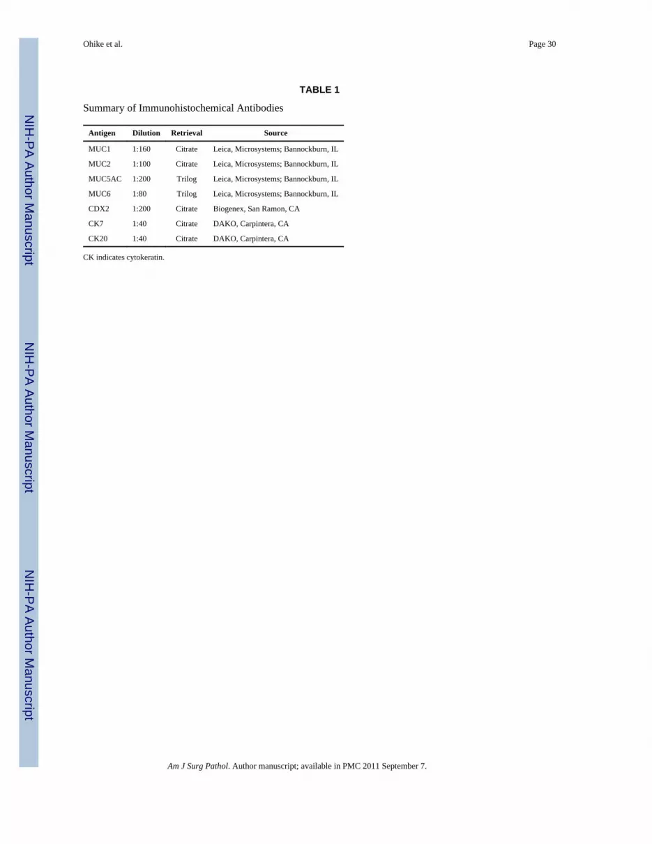
temperature. Between incubations, sections were washed with tris-buffered saline.Coverslipping was performed using the Tissue-Tek SCA coverslipper (Sakura Finetek USA,Inc, Torrance, CA). The negative (for which the primary antibody was replaced by tris-buffered saline) and positive controls were processed with the other slides in the study. Thedetailed specifications of the antibodies are provided in Table 1.
Evaluation of Immunohistochemistry—The percent of cells showing membranous(MUC1), cytoplasmic (MUC2, MUC5AC, MUC6, CK7, and CK20), and nuclear (CDX2)labeling were recorded. As is customary in most studies, labeling in less than 10% of thecells was designated as “negligible/negative” expression. Labeling in 10% to 50% of tumorcells was regarded as “focal positive,” whereas greater than 50% was considered “diffusepositive.”
Statistical Analysis—Patient and tumor characteristics were analyzed by an unpairedStudent t test or χ2 tests. Overall survival was analyzed using the Kaplan-Meier method anddifferences among groups were assessed by log-rank test. A Cox proportional hazardregression (version 2.9.1; open source statistical software) was used to identify independentfactors associated with postresection survival. All tests were 2-sided, and statisticalsignificance was defined as P value less than 0.05.
RESULTSGeneral Characteristics
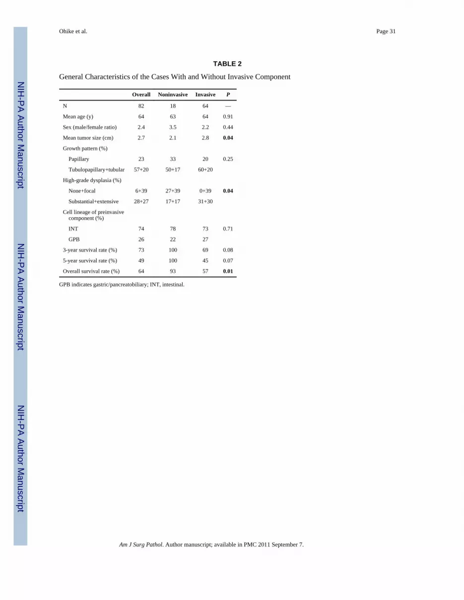
Eighty-two cases fulfilled the criteria described for IAPN (Textbox 1). The prevalenceamong ampullary glandular neoplasms was 33% and 5.5% among pancreatoduodenal/ampullary resection specimens. The mean age was 64 years (range: 27 to 85 y). Fifty-sevenpatients were male and 24 were female (ratio: 2.4). Main presentation symptoms wereavailable in 19 cases and included jaundice (19), weight loss (4), abdominal pain (4),nausea/vomiting (3), pruritis (2), and dark urine/light stool (2), and tarry stool (1). Imagingstudies (5 endoscopic retrograde cholangiopancreatography, 9 computed tomography, and 1magnetic resonance imaging) generally described dilated CBD and intra/extrahepatic ducts,ampullary, duodenal or pancreatic mass, and enlarged and/or irregular papillae. Thepreoperative diagnosis was available for 49 cases and was “ampullary mass” in 18 cases,“ampullary carcinoma” in 20, “duodenal carcinoma” in 4, “distal CBD carcinoma” in 3,“pancreatic carcinoma” in 1, and “pancreatic mass” in 3 cases (Table 2).
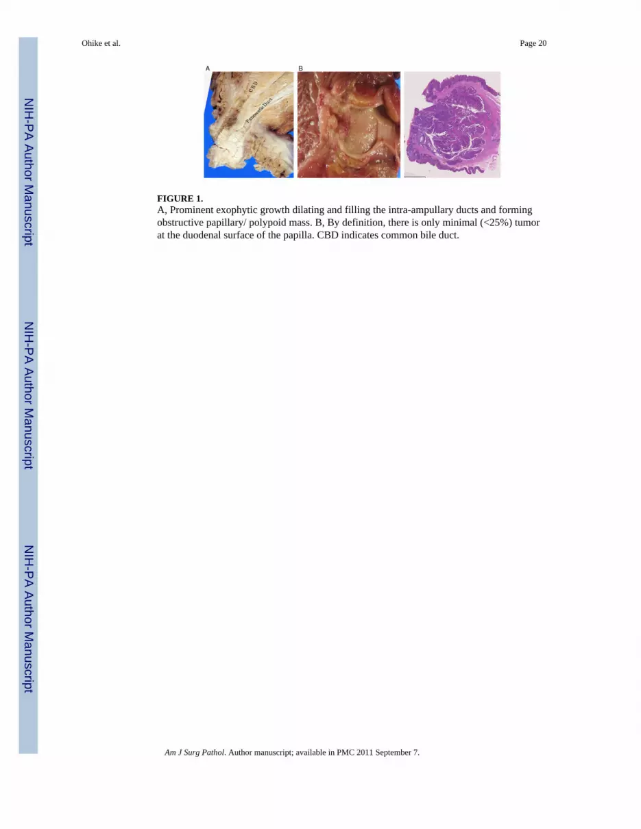
Macroscopic FindingsThese tumors were characterized by prominent exophytic growth within dilated intra-ampullary ducts and formation of obstructive papillary/polypoid masses, often withdilatation of the upstream biliary or pancreatic ducts (Fig. 1). The macroscopic patternmostly corresponded to what has been referred to as an “intramural protruding form,”57,63
although in rare cases there was minimal involvement of the duodenal surface of the papillaof Vater, thus representing the “exposed protruding form” or “ulcerating form.” Bydefinition, those cases with more prominent involvement of the papilla were excluded.Mucin hypersecretion was recognized in 7 cases (8.5%). The mean overall tumor size was2.7 cm (range: 0.8 to 6.7 cm). Although 2 preinvasive tumors were less than 1 cm (0.8 and0.9 cm), they were included in this study because they were easily distinguished from theflat lesions by their compact exophytic growth (Fig. 1) and their clear-cut delineation fromthe surrounding tissue.
Ohike et al. Page 6
Am J Surg Pathol. Author manuscript; available in PMC 2011 September 7.
NIH
-PA Author Manuscript
NIH
-PA Author Manuscript
NIH
-PA Author Manuscript
Microscopic FindingsThese lesions were characterized by well-organized, back to back epithelial units, either intubular (Fig. 2A) or papillary (Fig. 2B) architecture or both, with minimal or no interveningstroma between the units, similar to other preinvasive neoplasms. The base of the lesionswas sharply demarcated; however, the dysplastic process often showed complexity becauseof the involvement of the tributary glands comprising the ampullary ductules. This createdan architectural complexity and a pseudoinvasive pattern, which, at times, was difficult todistinguish from true invasion. In 38 cases (46%), the exophytic (preinvasive) lesionextended by pagetoid spread to the duodenal surface of the papilla of Vater, but bydefinition this duodenal lumen-facing component comprised less than 25% of the tumor.
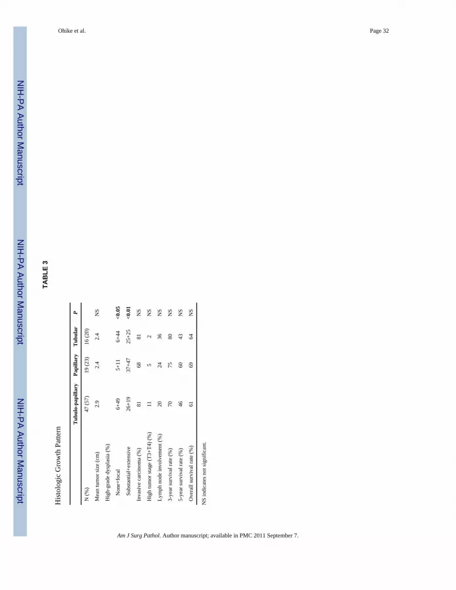
Histologic Growth PatternForty-seven cases (57%) had a tubulopapillary configuration (each component was >25% ofthe lesion), (Tables 2, 3), whereas 19 (23%) were predominantly (>75%) papillary (orvillous), and 16 (20%) were predominantly tubular. The mean tumor size did not seem todiffer between these groups: 2.4 cm in the papillary and tubular groups and 2.9 cm in thetubulopapillary group (Table 3).
Papillary examples had a higher tendency for HGD than the tubular and tubulopapillarygroups (P<0.05). The extent of HGD in the papillary group was focal in 11%, substantial in37%, and extensive in 47% of cases; these figures were 49%, 26%, and 19% fortubulopapillary, and 44%, 25%, and 25% for tubular examples. However, the prevalence ofan associated invasive carcinoma was lower in the papillary group (68%) than intubulopapillary (81%) or tubular (81%) groups. There were no significant differences inprognostic factors such as tumor stage, lymph node involvement, or 3 and 5-year survivalrates between these groups.
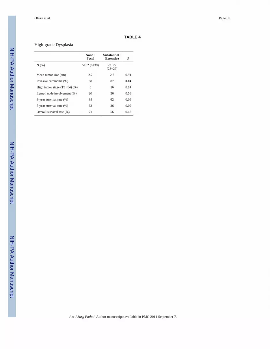
DysplasiaFive cases (6%) had no HGD, 32 (39%) had focal, 23 (28%) had substantial, and 22 cases(27%) had extensive HGD (Fig. 3). The prevalence of an associated invasive carcinoma forthese groups was 0%, 76%, 87%, and 86%. In addition, the 3 and 5-year survival rates of theformer 2 groups (focal or no HGD) seemed to be higher than those of the latter 2 groups(substantial or extensive HGD), but the difference was not statistically significant (P = 0.09)(Table 4).
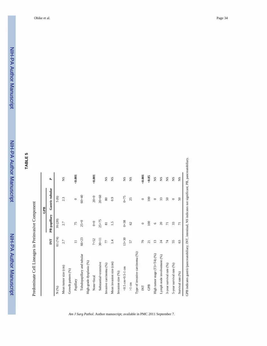
Histomorphologic Subtyping Based on Cell LineageUsing an approach that acknowledged any existing pattern of cell-lineage morphology,almost half of the cases (37 cases; 45%) were found to have mixed patterns (at least somefoci of a secondary pattern, Fig. 4), whereas 35 (43%) were pure INT (Fig. 5) and 10 caseswere (12%) pure GPB (Fig. 6). When evaluated with the forcedbinary classificationapproach, the predominant pattern was INT (Fig. 5) in 61 cases (74%) and GPB (Fig. 6) in21 cases (26%) (Table 5).
On the basis of the combination of predominate cell-lineage morphology and growth pattern,the cases were classified into the following categories:
INT-type (61 Cases)—The majority of INT-type cases were similar to conventionalcolonic/duodenal tubular adenomas consisting of relatively simple villous or tubularglandular units, lined by tall columnar INT-type cells with pseudostratified cigar-shapednuclei (Fig. 4A). Occasionally, cribriform architecture was observed. The apical cytoplasmwas usually densely chromophilic, but was pale or clear with abundant apical mucin in 3
Ohike et al. Page 7
Am J Surg Pathol. Author manuscript; available in PMC 2011 September 7.
NIH
-PA Author Manuscript
NIH
-PA Author Manuscript
NIH
-PA Author Manuscript
cases. Goblet, brush border, and/or Paneth cells were prominent in some cases (Fig. 5B).Mucin hyperproduction into the lumen, akin to that seen in pancreatic IPMNs, was seen inonly 3 cases.
Forty (66%) of the INT-type cases were tubulopapillary, 14 (23%) were tubular, and 7(11%) were papillary (or villous). Importantly, 43% had secondary GPB cells (Fig. 4),which is unusual in conventional colonic/duodenal adenomas. Prevalence and proportion ofHGD in the INT group was significantly lower than that of the GPB group presented below.Seven percent of cases had no HGD and 52% had focal, whereas 30% had substantial, andonly 11% of cases had extensive HGD. An associated invasive carcinoma was identified in77% of INT cases. The type of invasive carcinoma was INT in 79% and GPB in 21% ofthese cases; the size of invasion was less than 0.5 cm in 13%, 0.5 to 1 cm in 30%, andgreater than 1 cm in 57% of cases.
GPB-type (21 Cases)—Two distinct subtypes of GPB could be discerned based oncytoarchitectural characteristics. The first was characterized by a papillary (75%) ortubulopapillary (25%) growth pattern and cytomorphology similar to that of PB-type IPMNs(Fig. 6A). The second GPB subtype was characterized by a striking tubular growth patternand an overall appearance of a pyloric gland adenoma (Fig. 6B).
PB-papillary subtype (16 cases): Almost all PB-papillary-type cases had complex arborizingpapillae. The cytologic features often showed transition to a more gastric phenotype (Fig.6A), which further supported their kinship with the gastric type and also justified theircombined classification. However, all had HGD/carcinoma in situ in the papillary areas. Inaddition, HGD was extensive in 75% and substantial in the remaining 25%; none of thecases were classified as focal. An associated invasive carcinoma was identified in 81% ofPB-papillary-type cases all of which were GPB-type, and tended to be larger (0% were <0.5cm, 38% were 0.5 to 1 cm, and 62% >1 cm) than invasions seen in INT-type or the gastric-tubular type discussed below.
Gastric-tubular subtype (5 cases): The gastric-tubular-type lesion was recognized as aseparate variant because of its highly distinctive morphology among lesions with GPBlineage. The lesions were characterized by a closely packed, uniform, tubular growth patternreminiscent of pyloric or Brunner glands. The glands were arranged back-to-back and linedby a single layer of cuboidal to columnar GPB-type cells with basally oriented nuclei andabundant apical mucinous cytoplasm (Fig. 6B). Two gastric-tubular-type cases formedpolypoid masses resembling the pyloric gland type of adenoma in that one was devoid ofHGD and the other exhibited transformation into arborized papillary areas with HGD. Theremaining 3 cases showed monotonous, small tubular proliferation with extensive HGD andformation of expansile masses which resembled intraductal tubular/tubulopapillarycarcinoma of the pancreas, although some with a more mucinous-appearing cytoplasm.Expansile proliferation replaced most of the residual ductal epithelium and made recognitionof the intraductal (duct-confined) nature of the process challenging. An associated invasivecarcinoma was present in 4 cases (80%) and all were of PB-type. Invasion, however, seemedto be relatively small at the time of diagnosis, compared with INT-type or PB-papillarysubtype, in that 3 (75%) were 0.5 to 1.0 cm, and only 1 (25%) was larger than 1 cm.
No significant difference in prognostic factors such as pathologic tumor (pT) stage, lymphnode involvement, or 3 and 5-year survival rates were identified among these subtypes.
Cases With an Associated Invasive Carcinoma (64 Cases)Sixty-four of 82 cases (78%) had an associated invasive carcinoma (64 of 230, 28%, of allinvasive carcinomas within the ampulla arose in association with IAPNs). The average age
Ohike et al. Page 8
Am J Surg Pathol. Author manuscript; available in PMC 2011 September 7.
NIH
-PA Author Manuscript
NIH
-PA Author Manuscript
NIH
-PA Author Manuscript
and sex ratio of the patients was similar in the invasive and noninvasive groups (64 vs. 63 y;the male/female ratio was 2.2 vs. 3.5, P = 0.91 and P = 0.44, respectively). The overall sizeof the tumor tended to be larger in the invasive group than in the noninvasive (2.8 vs. 2.1cm, respectively; P = 0.04). The size of invasion was less than 0.5 cm in 6 cases (9%), 0.5 to1 cm in 22 (34%), and greater than 1 cm in 36 cases (56%).
The histologic type of invasive carcinoma included 60 ordinary adenocarcinomas and 4mixed adenocarcinomas (2 with mucinous, 1 squamous, and 1 with neuroendocrinecomponents).
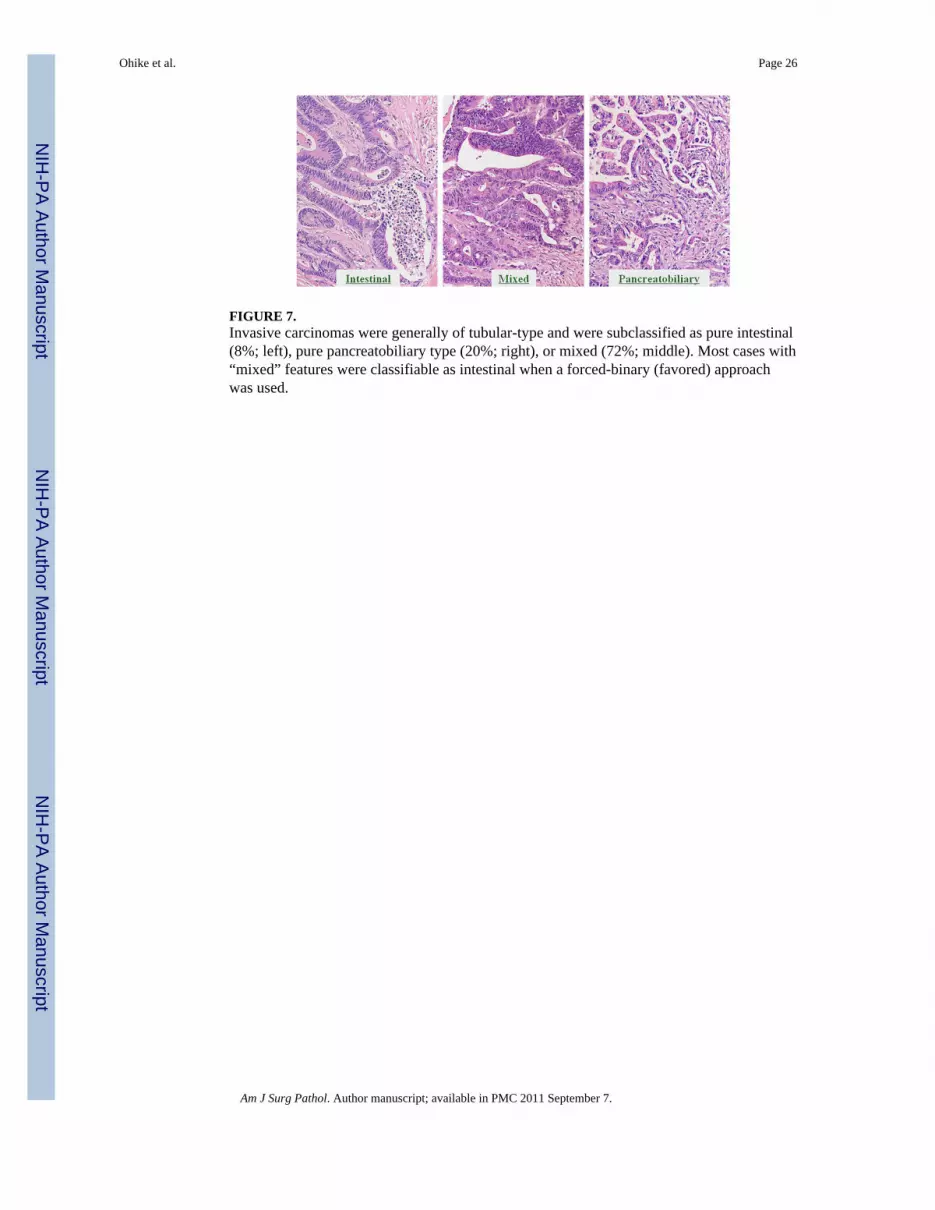
As for the cell-lineage morphology, associated invasive carcinomas were classified as pureINT-type in 5 cases (8%), mixed in 46 (72%), and pure PB-type in 13 cases (20%). On thebasis of the forced-binary approach (which eliminated the mixed category), however, thecases were finally divided into INT-type (37 cases; 58%) and PB-type (27 cases; 42%). Therepresentative histology of each type is shown in Figure 7.
In terms of preinvasive components, among the 61 preinvasive lesions of INT cell lineage,47 (77%) had an associated invasive carcinoma including 37 invasive lesions of INT celllineage and 10 invasive lesions of PB cell lineage. On the other hand, among the 21preinvasive lesions of GPB cell lineage, 17 (81%) had an associated invasive carcinoma allof which were of PB cell lineage (Table 5).
Among the 64 cases with an associated invasive carcinoma, the preinvasive component wasINT-type in 47 (73%) and GPB-type in 17 cases (27%). There was lineagetype concordancebetween preinvasive and invasive components in 54 cases, whereas 10 were discordant; all10 discordant cases had INT-type preinvasive neoplasms but PB-type invasion. All caseswith a GPB-type preinvasive component also had a PB-type invasive component.
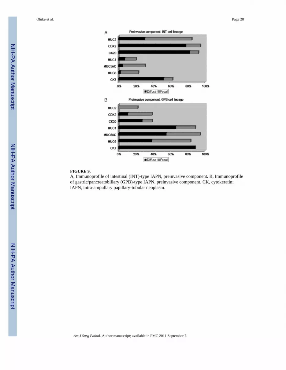
Immunophenotypic FeaturesPreinvasive Components—Fifty-nine cases were available for immunohistochemicalstaining. The preinvasive components of INT cell-lineage morphology (41 cases) showedimmunolabeling (diffuse+focal) for MUC2 (31%+54%) in 85% of the cases, 94% (78%+16%) for CDX2, 93% (83%+10%) for CK20, 21% (8%+13%) for MUC1, 31% (5%+26%)for MUC5AC, 24% (3%+21%) for MUC6, and 63% of cases (53%+10%) for CK7 (Figs. 8,9A).
Those with GPB cell-lineage morphology (18 cases) showed immunolabeling (diffuse+focal) for MUC2 in 22% (0%+22%) of the cases, 39% (11%+28%) for CDX2, 39% (28%+11%) for CK20, 89% (67%+22%) for MUC1, 95% (56%+39%) for MUC5AC, 83% (39%+44%) for MUC6, and 89% (89%+0%) of cases showed immunolabeling for CK7 (Figs. 8,9B).
The sensitivity and specificity for INT cell-lineage markers were as follows: MUC2, 85%and 78%; CDX2, 94% and 61%; and CK20, 93% and 61%. The sensitivity and specificityfor GPB cell-lineage markers were as follows: MUC1, 89% and 79%; MUC5AC, 95% and69%; and MUC6, 83% and 76%. CKs 7 and 20 were coexpressed in 53% of all casesavailable for immunohistochemical staining.
When the subsets of GPB type were analyzed separately, it was noted that all PB-papillarycases had at least focal MUC5AC, and all gastric-tubular subtype had at least focalexpression of MUC1.
Ohike et al. Page 9
Am J Surg Pathol. Author manuscript; available in PMC 2011 September 7.
NIH
-PA Author Manuscript
NIH
-PA Author Manuscript
NIH
-PA Author Manuscript
Invasive Components—Tissue from 24 invasive INT-type carcinomas was available forevaluation. Immunolabeling (diffuse+focal) was positive for the following percentage ofcases: MUC2 67% (25%+42%); CDX2 78% (52%+26%); CK20 83% (75%+8%); MUC146% (4%+42%); MUC5AC 59% (13%+46%); MUC6 17% (4%+13%); and CK7 67% (54%+13%).
Twenty-three PB-type invasive carcinomas were available for evaluation. Immunolabeling(diffuse+focal) was positive for the following percentage of cases: MUC2 17% (4%+13%);CDX2 34% (17%+17%); CK20 48% (35%+13%); MUC1 96% (83%+13%); MUC5AC61% (26%+35%); MUC6 39% (22%+17%); and CK7 83% (83%+0%).
For invasive carcinoma of the INT cell lineage, sensitivity and specificity results were asfollows: MUC2 67% and 83%; CDX2 78% and 66%; and CK20 83% and 52%. Sensitivityand specificity for invasive carcinoma of the PB cell lineage were as follows: MUC1 96%and 54%; MUC5AC 61% and 41%; MUC6 39%and 83%; and CK7 83% and 33%.
Prognosis (Clinical Outcome)Prognostic Stratifiers—Cases without invasion had a very good prognosis, as evidencedby 3 and 5-year survival rates (100% and 100%) significantly better than those cases with anassociated invasive carcinoma (69% and 45%, respectively) (Table 2). There weresignificant differences between these 2 groups in terms of tumor size (2.1 vs. 2.8 cm), andHGD proportion (no or focal HGD/substantial or extensive HGD 67%/33%, P = 0.04 vs.39%/61%, P = 0.04). Only 1 patient with noninvasive carcinoma died; he had substantialHGD/carcinoma in situ and died 85 months after undergoing a Whipple resection.
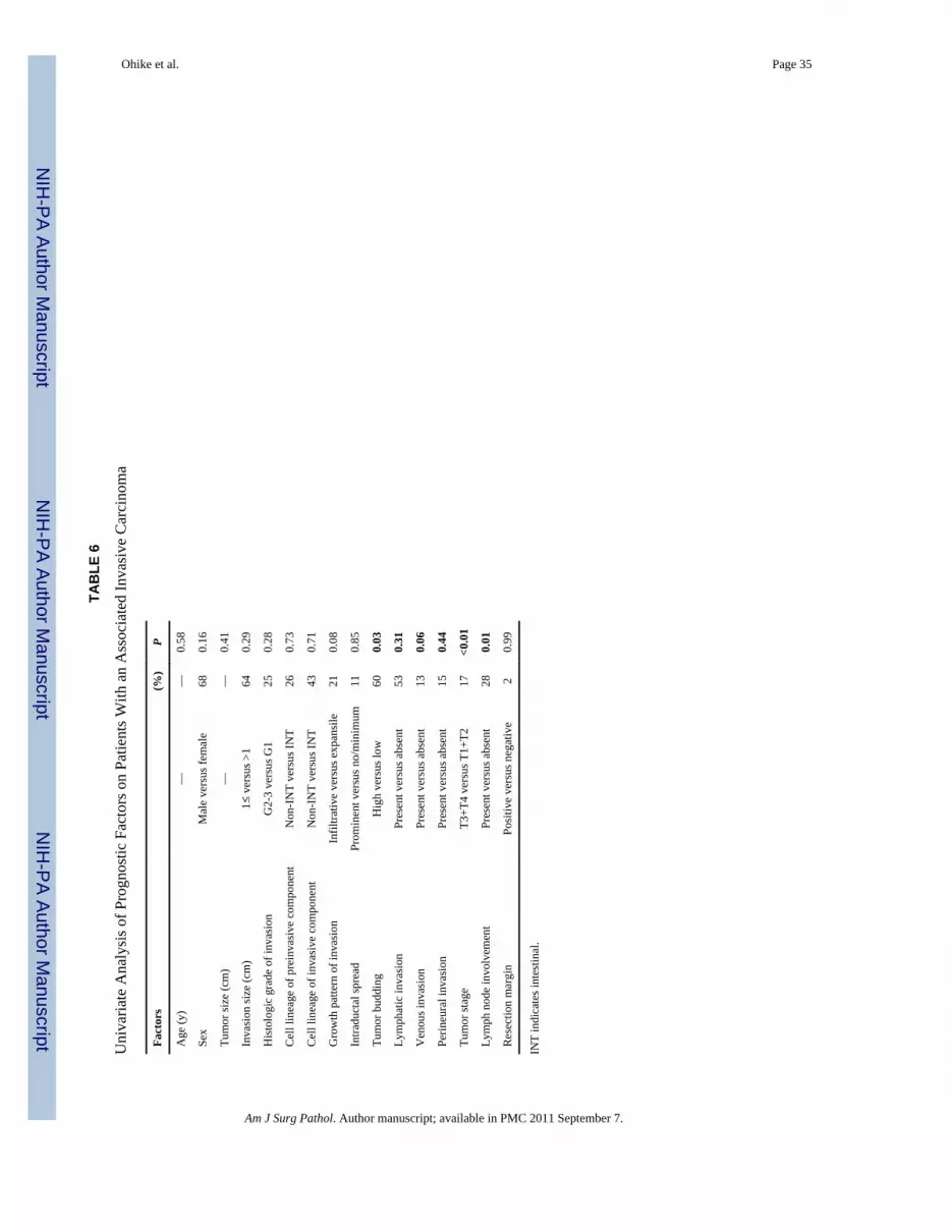
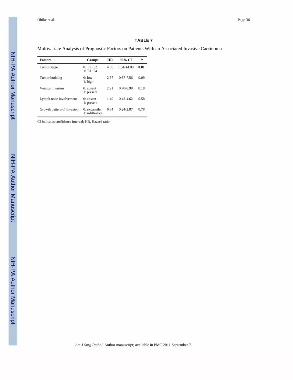
Univariate and multivariate analyses were performed on 53 cases with an associatedinvasive carcinoma for which all the parameters tested were available (eg, cases withincomplete lymph node dissection because of local excision, like ampullectomies, wereexcluded). In univariate analysis, pT stage (P<0.01), lymph node involvement (P = 0.01),and tumor budding (P = 0.03) were found to be the significant prognostic factors, whereasvenous invasion (P = 0.06) and pattern of invasion (infiltrative vs. expansile) alsoapproached statistical significance (P = 0.08) (Table 6). In a stepwise, multivariable Coxregression analysis which incorporated these 5 factors, pT stage (P = 0.01) was the onlysignificant independent factor related to prognosis, although tumor budding did approachstatistical significance (P = 0.09) (Table 7).
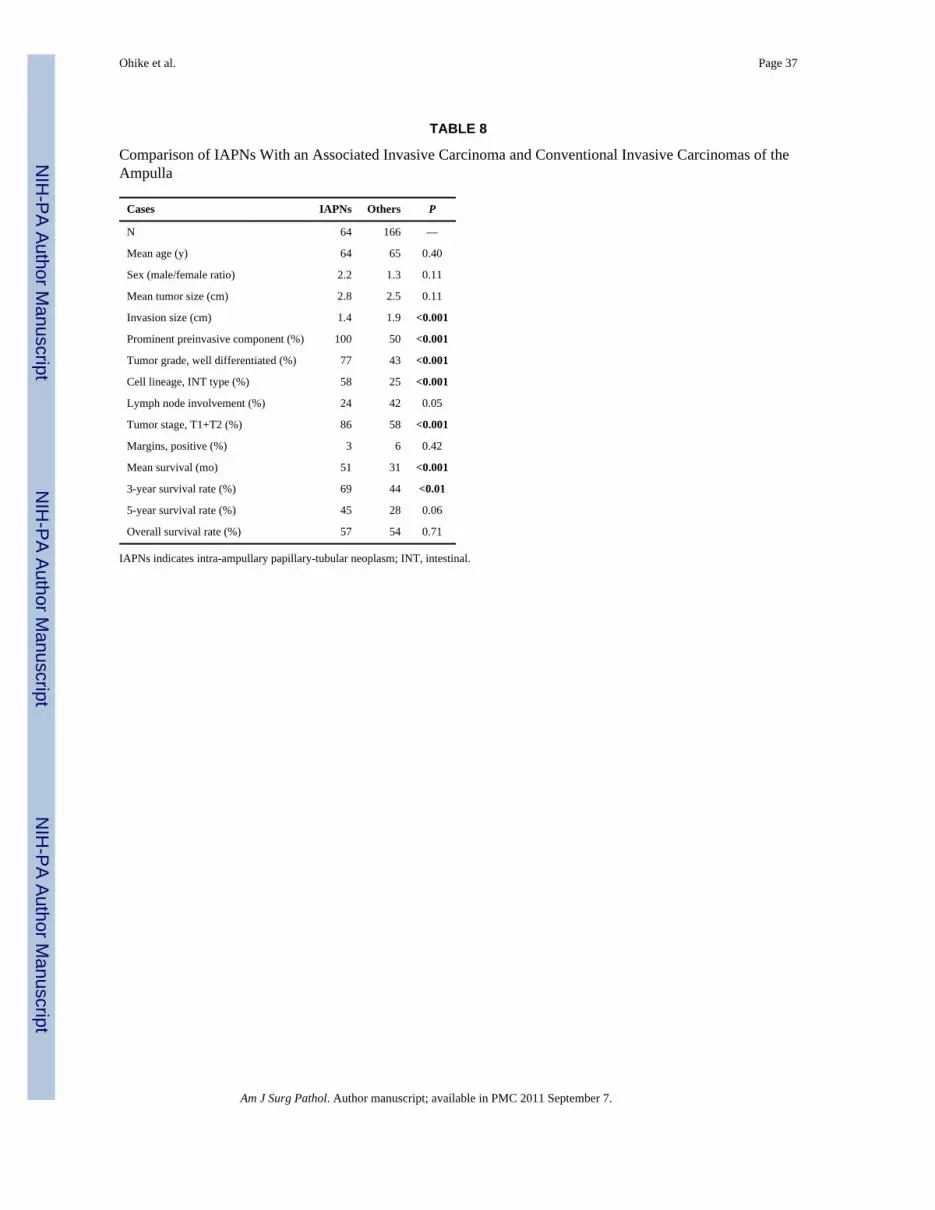
Comparison With Other Ampullary CarcinomasWhen IAPNs with an associated invasive carcinoma were compared with conventionalinvasive carcinomas of the ampulla, the mean tumor size was not significantly different (2.8cm vs. 2.5 cm), but there was a significant difference in the size of invasion (mean size ofinvasion 1.4 cm vs. 1.9 cm, P<0.001). Minimal invasion (<5 mm) was more common in theformer (9% vs. 2%). IAPNs with an associated invasive carcinoma also had significantlybetter tumor differentiation (P<0.001), were more frequently of INT cell lineage (P<0.001),had a lower tumor stage (P<0.001), and had less lymph node involvement (P = 0.05). Moreimportantly, cases of invasion associated with IAPNs had a significantly better prognosisthan conventional invasive carcinomas of the ampulla. The mean survival time was 51versus 31 months (P<0.001) and the 3-year survival rate was 69% versus 44% (P<0.01);although, the difference in 5-year survival rate (45% vs. 28%) did not reach statisticalsignificance (P = 0.06) (Table 8, Fig. 10).
Ohike et al. Page 10
Am J Surg Pathol. Author manuscript; available in PMC 2011 September 7.
NIH
-PA Author Manuscript
NIH
-PA Author Manuscript
NIH
-PA Author Manuscript
DISCUSSIONDefinition and Delineation From Similar Neoplasms
The tumor-forming intraepithelial neoplasms analyzed in this study are remarkablyanalogous to IPMNs and ITPNs of the pancreas and to biliary IPNs.2,3,25,26,29,64,69 Theirstriking similarities with these neoplasms warrant their classification in a parallel category.As with their counterparts in the neighboring sites, these neoplasms are characterized byexophytic (papillary and/or polypoidtubular) growth, variable cellular lineage (INT or GBP),and a spectrum of dysplastic change (adenoma-carcinoma sequence). Until now, they havebeen classified as conventional adenocarcinomas of the ampulla, or included in studies on“adenomas” of the duodenum, which may involve the duodenal surface of the papilla ofVater, and classified as ampullary. However, it is clear that they must be distinguished fromthe former, because, as is now widely accepted, invasive carcinomas arising in associationwith mass-forming preinvasive neoplasms (eg, invasive carcinomas in IPMNs) havedistinctive characteristics, including different biology and clinical behavior. Theseneoplasms also ought to be delineated from the conventional INT-type duodenal adenomasthat involve the duodenal surface at the Vaterian region. Although there are varioussimilarities between these entities, it is necessary to classify the intra-ampullary onesseparately as is carried out for the mass-forming preinvasive neoplasms in the PB tract,which are classified based on their specific site of origin, and furthermore, it is safe toassume that the internal aspect of the ampulla (a transitional region with various distinctivehistomorphologic characteristics) confers specific properties to the tumors. Indeed, in thisstudy, these neoplasms were found to have various site-specific characteristics, as discussedbelow.
TerminologyHerein, we provisionally propose to designate these neoplasms as “intra-ampullarypapillary-tubular neoplasm.” The term “intra-ampullary” is proposed to indicate both thelocalized and preinvasive nature of these neoplasms. Their exophytic (mass-forming) nature,in contrast, is highlighted by the name “papillarytubular,” which emphasizes the fact thatthese tumors can have either papillary or tubular or both configurations, in a mixture. Thename papillary-tubular is favored over tubulopapillary (or “tubulovillous”), because, thelatter (1) is a term that has been specific to mixed-type neoplasms in the gastrointestinaltract, whereas, with IAPNs, the mixed type is common but some cases are purely papillaryor purely tubular; (2) may give the impression that these neoplasms are more commonlytubular than papillary, but in fact the papillary pattern is more common; and (3) is now aterm used in the category designation for a specific pancreatic tumor type, namely ITPNs26
(a designation also advocated in the upcoming WHO classification). Only a smallpercentage of IAPNs display the features of pancreatic ITPNs. Finally, the possibility ofincorporating the word “mucinous” in the category designation was considered butdismissed, because, this descriptor was recently dropped from the nomenclature of biliaryneoplasms because of the fact that, like IAPNs, they often produce only minimal mucin.This is in contrast with pancreatic IPMNs, which are often characterized by profuse mucinproduction.
IncidenceIAPNs are relatively rare. They constitute 33% (82 of 248 cases) of primary ampullarytumors and 5.5% (82 of 1480 cases) of pancreatoduodenectomies/ampullectomies withneoplasm. The frequency of a preinvasive component in ampullary cancers is highlyvariable in the literature, presumably because of marked definitional differences. In somestudies, it was reported to be up to 90%. In this study, one third of the cases with invasivecarcinoma in the ampulla were associated with an IAPN component. This percentage is
Ohike et al. Page 11
Am J Surg Pathol. Author manuscript; available in PMC 2011 September 7.
NIH
-PA Author Manuscript
NIH
-PA Author Manuscript
NIH
-PA Author Manuscript
higher than in the pancreas where IPMNs have been implicated in 2%30 to 10%47 ofinvasive carcinomas, or in the gallbladder where we recently found 10% of invasive cancersto have an associated tumoral intraepithelial neoplasia20 (reportedly 3% to 18% in theliterature).
Clinicopathologic CharacteristicsThe mean age at diagnosis, 64 years, is similar to that of patients with other cancers of theampullopancreatobiliary region.30 As in conventional carcinomas of the ampullary region,they occur predominantly in males (male/female ratio: 2.4 vs. 1.3). The mean tumor size isalso similar to that of the resected carcinomas seen in pancreatoduodenectomies: 2.8 cm (0.8to 6.7 cm; vs. 3 cm in other carcinomas). Not surprisingly, the patients often present withjaundice.
Macroscopically, most IAPNs, by definition, have minimal exophytic growth at theduodenal surface aspect of the papilla of Vater. However, they sometimes form a mucosalprotuberance in the duodenal lumen because of the underlying proliferative process, whichfills the intraampullary/ductal lumen. The duodenal orifice of the ampulla is often wide,irregular, and occasionally ulcerated. On cut sections of ampulla, IAPNs are characterizedby polypoid or papillary (granular-exophytic) lesions, which fill the ampullary channel (orthe intra-ampullary tips of both the CBD and pancreatic duct).
Microscopically, there is a variable degree of papillary and tubular configuration. About aquarter of the lesions are predominantly (>75%) composed of papillary elements, anotherquarter are tubular; however, more than half of the cases exhibit a substantial mixture of the2 growth patterns (ie, tubulopapillary). The frequency of the mixed pattern may partially beattributable to the complexity of the ampullary mucosa, which contains numerous tributaryglands, which impart a tubular architecture to the process when involved by the pagetoidspread of IAPN cells. The significance of the growth patterns needs further analysis, becausethe incidence and amount of HGD tended to be higher in those cases with papillaryconfiguration although did not otherwise seem to have statistically significant correlationwith either incidence of invasion or overall prognosis.
IAPNs display a spectrum of dysplasia (neoplastic transformation) not only within theentity, but also commonly within a given patient. More than 80% of the cases exhibit amixture of low and high-grade dysplastic foci within the same lesion. The overall incidenceof HGD is very high; only a small minority (6%) of cases is entirely devoid of HGD. Inabout one third of the cases, HGD is focal, in another third, substantial (25% to 50% of thelesion), and yet, in about a quarter, it is extensive. The existence and amount of HGD seemto trend toward higher incidence of invasive carcinoma and more aggressive behavior;however, this association did not reach statistical significance in this study. Of note, all 5cases, which lacked HGD also lacked invasive carcinoma. This very high incidence of HGDis intriguing especially considering the relatively early stage detection of these lesions. Thissuggests that once the neoplastic transformation begins in the ampullary epithelium, itprogresses rather rapidly. This may be attributable to either some inherent vulnerability ofthis mucosa or its ready exposure to different milieu with its potential instigators includingpancreatic secretions, bile, and duodenal secretions.
Like their counterparts in the pancreatic and biliary tract, IAPNs exhibit a spectrum of celllineages that recapitulate those found in different compartments of the gastrointestinal tract.Interestingly, in about half of the cases, a mixture of these cell lineages is seen incoexistence, which may not be surprising considering the transitional nature of the ampulla.This high incidence of mixed lineage also distinguishes IAPNs from the conventional INT-
Ohike et al. Page 12
Am J Surg Pathol. Author manuscript; available in PMC 2011 September 7.
NIH
-PA Author Manuscript
NIH
-PA Author Manuscript
NIH
-PA Author Manuscript
type adenomas that occur at the duodenal surface of the papilla of Vater, which areuniformly of INT-type and often show different molecular events.51
Although mixed lineage was common when neoplasms were evaluated using a purist’sapproach, when a forced-binary approach based on the predominant pattern was used, themajority (74%) of IAPN cases could be designed INT type (closely resembling pancreaticINT-type IPMNs or colonic adenomas than GPB lesions), whereas the remaining 26% wereplaced into the GPB category. However, many cases placed into the INT category exhibitedsubtle, often hybrid features different from ordinary colonic adenomas. The GPB categorycould be further subdivided into 2 groups (1) those with a prominent papillary configurationresembling PB-type IPMNs, and (2) those which are predominantly tubular with pyloricgland adenoma-type features. Although the prevalence of extensive HGD was fairly low inINT cases (11%), it was fairly common (75%) in PB-papillary cases. The associatedinvasive carcinoma also tended to be smaller in the INT-type. The gastric tubular-type caseswere rare (6%) and more difficult to characterize. Interestingly, 4 of 5 gastric-tubular-typecases in this study had an associated invasive carcinoma but 3 of these were smaller than 1cm.
Immunohistochemical markers used in IAPNs do not have the same degree of reliability fordistinguishing cell lineages as they do, for example, in pancreatic IPMNs.8,22,43,53 This isnot surprising when the hybrid and transitional nature of the ampulla, as well as thefrequency of mixed phenotypes at the histologic level are considered. Even the most reliablemarker for INT differentiation in IAPNs, MUC2, was found to have sensitivity andspecificity of only 85% and 78%, respectively, whereas CDX2 was more sensitive butentirely lacked specificity (90% and 61%). For the GPB lineage, markers that had somedegree of sensitivity and specificity were MUC1 (89% and 79%) and MUC6 (83% and76%). Moreover, the CK profile was entirely nondiscriminatory, in that, CK7 and CK20were coexpressed in more than half of the cases. More importantly, CK7, which is regardedas a good marker of GPB differentiation was expressed in a high proportion (63%) of INTcases, and CK20, which is generally considered a good marker of the INT phenotype, wasexpressed in a substantial number (39%) of GPB. This divergence from the conventional,expected profile can be taken as further evidence of the transitional nature of IAPNs andtheir pronounced ability to form hybrid phenotypes.
Invasive Carcinomas Associated With IAPNsIAPNs commonly (78%) have an associated invasive carcinoma at the time of diagnosis.However, in almost half of these, the invasive carcinoma is less than 1 cm. Invasivecarcinoma is more prone to develop in larger IAPNs (mean size was 2.8 cm in invasive vs.2.1 cm in noninvasive cases, P = 0.04). All cases that have an associated invasive carcinomahave at least focal HGD. All 5 cases that lacked HGD also lacked an associated invasivecarcinoma; however, overall, the amount of HGD does not seem to correlate significantlywith invasion. There does not seem to be any association between propensity for invasionand the configuration (papillary vs. tubular) of the preinvasive neoplasm.
Invasive carcinomas that arise in IAPNs are mostly tubule-forming (glandular-type)adenocarcinomas that commonly (72%) show INT/PB hybrid features. This furthercorroborates the transitional nature of this area and its capacity to form different lineages.However, when a forced-binary classification based on the predominant (favored) patternwas used, 58% of invasive carcinomas in IAPN become classifiable as INT and 42% as PB.The cell lineage of an associated invasive carcinoma often but not always parallels that ofthe noninvasive component; in 84% (54 of 64), there was a correlation between the 2components and in 16% (10 of 64), there was a discrepancy. The discrepancy occurred only
Ohike et al. Page 13
Am J Surg Pathol. Author manuscript; available in PMC 2011 September 7.
NIH
-PA Author Manuscript
NIH
-PA Author Manuscript
NIH
-PA Author Manuscript
in INT-type IAPN, of which 21% (10 of 47) led to PB-type invasive carcinoma. All invasivecarcinomas that arose from GBP-type IAPNs were of PB-type.
Biologic Behavior and Clinical Outcome ParametersThe prognosis for IAPNs when both noninvasive and invasive cases are considered, issignificantly better than that of other invasive carcinomas of the ampulla, and isincomparably better than pancreatic ductal adenocarcinoma. Not surprisingly, noninvasiveexamples have a far better prognosis: the 3 and 5-year survival rates are 100%. In this study,the only patient from the noninvasive group who died of disease had extensive HGD anddied 8 years after undergoing a pancreatoduodenectomy. The survival rate for patients withinvasive IAPN (mean length of survival, 51 months, and the 3-year survival rate, 69%), isnot quite as good as the noninvasive cases but is nevertheless still better significantly thanthat of other invasive carcinomas of the ampulla without an accompanying IAPN component(mean length of survival is 31 months and the 3-year survival rate is 44%; P<0.01), althoughthe difference in survival rate at 5 years (45% vs. 28%) did not reach statistical significancein this study (P = 0.06), which raises suspicion that the gap between the 2 may diminish inlong-term follow-up. The survival advantage of IAPN-associated cases over non-IAPNassociated ones is partly attributable, not surprisingly, to early detection of the invasivecomponent tagged by the noninvasive (IAPN) process, including smaller size of invasion(1.4 vs. 1.9), more early-stage cancers (T1/2: 86% vs. 58%), and lower incidence of lymphnode metastasis (24% vs. 42%).
Within the group of invasive IAPNs, the pathologic parameters that correlated withprognosis in univariate analysis and thus are suggested to be included in surgical pathologyreports were as follows: stage (pT-stage and N-status), tumor budding, venous invasion, andinfiltrative versus expansile growth. Among these, the T-stage seemed to be the onlyindependent prognostic parameter, whereas budding approached the level of independence.
CONCLUSIONSIn conclusion, mass-forming preinvasive neoplasms occurring within the ampulla havemany similarities to biliary and pancreatic papillary, intraductal papillarymucinous, andtubulopapillary neoplasms, in that they exhibit a spectrum of dysplastic change, variableconfiguration, and different cell lineages, often in a mixture. We believe these neoplasmswarrant classification under a unified category for which we propose the descriptive nameIAPN. IAPNs are indolent, and have a better overall prognosis for invasive examples thanother invasive cancers which arose in this region. However, IAPNs associated with invasivecarcinoma often have a malignant clinical course, albeit not as aggressive as of those that areunaccompanied by IAPN.
AcknowledgmentsThe authors thank Leslie Ducato and Rhonda Everett for their assistance in the preparation of this manuscript, andAhnon Milham for her critical review of the article.
Supported in part by the National Cancer Institute Specialized Program in Research Excellence (SPORE)CA101936 in Pancreas Cancer (PAR-02-068) and in part by the Georgia Cancer Coalition Distinguished CancerClinicians and Scientists Program.
REFERENCES1. Adsay, NV.; Klöppel, G.; Fukushima, N., et al. Intraductal neoplasms of the exocrine pancreas. In:
Bosman, FT.; Carneiro, F.; Hruban, RH., et al., editors. World Health Organization Classification ofTumours of the Digestive System. WHO Press; Geneva: in press
Ohike et al. Page 14
Am J Surg Pathol. Author manuscript; available in PMC 2011 September 7.
NIH
-PA Author Manuscript
NIH
-PA Author Manuscript
NIH
-PA Author Manuscript
2. Abraham SC, Lee JH, Boitnott JK, et al. Microsatellite instability in intraductal papillary neoplasmsof the biliary tract. Mod Pathol. 2002; 15:1309–1317. [PubMed: 12481012]
3. Abraham SC, Lee JH, Hruban RH, et al. Molecular and immunohistochemical analysis ofintraductal papillary neoplasms of the biliary tract. Hum Pathol. 2003; 34:902–910. [PubMed:14562286]
4. Adsay, NV.; Klimstra, DS. Benign and malignant tumors of the gallbladder and extrahepatic biliarytract. In: Odze, RD.; Goldblum, JR., editors. Surgical Pathology of the GI tract, Liver, Biliary Tract,and Pancreas. Elsevier; Philadelphia: 2009. p. 845-875.
5. Adsay NV, Adair CF, Heffess CS, et al. Intraductal oncocytic papillary neoplasms of the pancreas.Am J Surg Pathol. 1996; 20:980–994. [PubMed: 8712298]
6. Adsay NV, Merati K, Andea A, et al. The dichotomy in the preinvasive neoplasia to invasivecarcinoma sequence in the pancreas: differential expression of MUC1 and MUC2 supports theexistence of two separate pathways of carcinogenesis. Mod Pathol. 2002; 15:1087–1095. [PubMed:12379756]
7. Adsay NV, Merati K, Nassar H, et al. Pathogenesis of colloid (pure mucinous) carcinoma ofexocrine organs: coupling of gel-forming mucin (MUC2) production with altered cell polarity andabnormal cell-stroma interaction may be the key factor in the morphogenesis and indolent behaviorof colloid carcinoma in the breast and pancreas. Am J Surg Pathol. 2003; 27:571–578. [PubMed:12717243]
8. Adsay NV, Merati K, Basturk O, et al. Pathologically and biologically distinct types of epitheliumin intraductal papillary mucinous neoplasms: delineation of an “intestinal” pathway ofcarcinogenesis in the pancreas. Am J Surg Pathol. 2004; 28:839–848. [PubMed: 15223952]
9. Albores-Saavedra, J.; Henson, DE.; Klimstra, DS. Atlas of Tumor Pathology. 3rd ed.. Armed ForcesInstitute of Pathology; Washington DC: 2000. Tumors of the gallbladder, extrahepatic bile ducts,and ampulla of Vater; p. 245-258.
10. Albores-Saavedra J, Murakata L, Krueger JE, et al. Noninvasive and minimally invasive papillarycarcinomas of the extrahepatic bile ducts. Cancer. 2000; 89:508–515. [PubMed: 10931449]
11. Albores-Saavedra J, Schwartz AM, Batich K, et al. Cancers of the ampulla of vater: demographics,morphology, and survival based on 5625 cases from the SEER program. J Surg Oncol. 2009;100:598–605. [PubMed: 19697352]
12. Alexander JR, Andrews JM, Buchi KN, et al. High prevalence of adenomatous polyps of theduodenal papilla in familial adenomatous polyposis. Dig Dis Sci. 1989; 34:167–170. [PubMed:2536604]
13. Bal A, Joshi K, Vaiphei K, et al. Primary duodenal neoplasms: a retrospective clinico-pathologicalanalysis. World J Gastroenterol. 2007; 13:1108–1111. [PubMed: 17373748]
15. Basturk, O.; Farris, AB.; Adsay, NV. Immunohistology of pancreas, gallbladder, extrahepatic bileducts, ampulla and liver. In: Dabbs, D., editor. Diagnostic Immunohistochemistry. 3rd ed..Saunders Elsevier; Philadelphia: 2010. p. 541-592.
16. Basturk O, Khayyata S, Klimstra DS, et al. Preferential expression of MUC6 in oncocytic andpancreatobiliary types of intraductal papillary neoplasms highlights a pyloro-pancreatic pathway inpancreatic carcinogenesis, distinct from the intestinal pathway. Am J Surg Pathol. 2010; 34:364–370. [PubMed: 20139757]
17. Cathro HP, Stoler MH. Expression of cytokeratins 7 and 20 in ovarian neoplasia. Am J Clin Pathol.2002; 117:944–951. [PubMed: 12047147]
18. Cavallini M, Cavaniglia D, Felicioni F, et al. Large periampullary villous tumor of the duodenum.J Hepatobiliary Pancreat Surg. 2007; 14:526–528. [PubMed: 17909726]
19. Chu P, Wu E, Weiss LM. Cytokeratin 7 and cytokeratin 20 expression in epithelial neoplasms: asurvey of 435 cases. Mod Pathol. 2000; 13:962–972. [PubMed: 11007036]
20. Dursun N, Roa JC, Tapia O, et al. Intravesicular papillary-tubular neoplasm (IVPN) as a unifyingcategory for mass formingpreinvasive neoplasms of the gallbladder: an analysis of 87 cases. ModPathol. 2010; 23:144A. [PubMed: 19838162]
Ohike et al. Page 15
Am J Surg Pathol. Author manuscript; available in PMC 2011 September 7.
NIH
-PA Author Manuscript
NIH
-PA Author Manuscript
NIH
-PA Author Manuscript
21. Duval JV, Savas L, Banner BF. Expression of cytokeratins 7 and 20 in carcinomas of theextrahepatic biliary tract, pancreas, and gallbladder. Arch Pathol Lab Med. 2000; 124:1196–1200.[PubMed: 10923083]
22. Furukawa T, Kloppel G, Adsay NV, et al. Classification of types of intraductal papillary-mucinousneoplasm of the pancreas: a consensus study. Virchows Arch. 2005; 447:794–799. [PubMed:16088402]
23. Goldstein NS, Bassi D. Cytokeratins 7, 17, and 20 reactivity in pancreatic and ampulla of vateradenocarcinomas. Percentage of positivity and distribution is affected by the cut-point threshold.Am J Clin Pathol. 2001; 115:695–702. [PubMed: 11345833]
24. Hartenfels IM, Dukat A, Burg J, et al. Adenomas of Vater’s ampulla and of the duodenum,presentation of diagnosis and therapy by endoscopic interventional and surgical methods. Chirurg.2002; 73:235–240. [PubMed: 11963497]
25. Hruban RH, Takaori K, Klimstra DS, et al. An illustrated consensus on the classification ofpancreatic intraepithelial neoplasia and intraductal papillary mucinous neoplasms. Am J SurgPathol. 2004; 28:977–987. [PubMed: 15252303]
26. Hruban, RH.; Pitman, MB.; Klimstra, DS. AFIP Atlas of Tumor Pathology. American Registry ofPathology; Washington, DC: 2007. Tumors of the Pancreas; p. 75-110.4th series, Fascicle 6
27. Ji H, Isacson C, Seidman JD, et al. Cytokeratins 7 and 20, Dpc4, and MUC5AC in the distinctionof metastatic mucinous carcinomas in the ovary from primary ovarian mucinous tumors: Dpc4assists in identifying metastatic pancreatic carcinomas. Int J Gynecol Pathol. 2002; 21:391–400.[PubMed: 12352188]
28. Kim HJ, Kim MH, Lee SK, et al. Mucin-hypersecreting bile duct tumor characterized by a strikinghomology with an intraductal papillary mucinous tumor (IPMT) of the pancreas. Endoscopy.2000; 32:389–393. [PubMed: 10817178]
29. Klimstra, DS.; Adsay, NV. Tumors of the pancreas and ampulla vater. In: Odze, RD.; Goldblum,JR., editors. Surgical Pathology of the GI tract, Liver, Biliary Tract, and Pancreas. Elsevier;Philadelphia: 2009. p. 845-876.
30. Klimstra, DS.; Adsay, NV. Tumors of the pancreas and ampulla of vater. In: Odze, RD.;Goldblum, JR., editors. Surgical Pathology of the GI tract, Liver, Biliary Tract, and Pancreas.Elsevier; Philadelphia: 2009. p. 909-962.
31. Klimstra DS, Adsay NV, Dhall D, et al. Intraductal tubular carcinoma of the pancreas:clinicopathologic and immunohistochemical analysis of 18 cases. Mod Pathol. 2007; 20:285A.
32. Kloppel G, Kosmahl M. Is the intraductal papillary mucinous neoplasia of the biliary tract acounterpart of pancreatic papillary mucinous neoplasm? J Hepatol. 2006; 44:249–250. [PubMed:16360969]
33. Kloppel, G.; Solcia, E.; Longnecker, DS., et al. Histologic Typing of Tumors of the ExocrinePancreas. Springer; Geneva: 1996.
34. Lee MJ, Lee HS, Kim WH, et al. Expression of mucins and cytokeratins in primary carcinomas ofthe digestive system. Mod Pathol. 2003; 16:403–410. [PubMed: 12748245]
35. Levi E, Klimstra DS, Andea A, et al. MUC1 and MUC2 in pancreatic neoplasia. J Clin Pathol.2004; 57:456–462. [PubMed: 15113850]
36. Li MK, Folpe AL. CDX-2, a new marker for adenocarcinoma of gastrointestinal origin. Adv AnatPathol. 2004; 11:101–105. [PubMed: 15090846]
37. Lim JH, Yi CA, Lim HK, et al. Radiological spectrum of intraductal papillary tumors of the bileducts. Korean J Radiol. 2002; 3:57–63. [PubMed: 11919480]
38. Lim JH, Jang KT, Rhim H, et al. Biliary cystic intraductal papillary mucinous tumor andcystadenoma/cystadenocarcinoma: differentiation by CT. Abdom Imaging. 2007; 32:644–651.[PubMed: 17437076]
39. Longnecker, DS.; Adler, G.; Hruban, RH., et al. Intraductal papillary mucinous neoplasms of thepancreas. In: Hamilton, SR.; Aaltonen, LA., editors. World Health Organization Classification ofTumours. Pathology and Genetics of Tumours of the Digestive System. IARC Press; Lyon: 2000.p. 237-243.
40. Luttges J, Zamboni G, Longnecker D, et al. The immunohistochemical mucin expression patterndistinguishes different types of intraductal papillary mucinous neoplasms of the pancreas and
Ohike et al. Page 16
Am J Surg Pathol. Author manuscript; available in PMC 2011 September 7.
NIH
-PA Author Manuscript
NIH
-PA Author Manuscript
NIH
-PA Author Manuscript
determines their relationship to mucinous noncystic carcinoma and ductal adenocarcinoma. Am JSurg Pathol. 2001; 25:942–948. [PubMed: 11420467]
41. Moskaluk CA, Zhang H, Powell SM, et al. Cdx2 protein expression in normal and malignanthuman tissues: an immunohistochemical survey using tissue microarrays. Mod Pathol. 2003;16:913–919. [PubMed: 13679455]
42. Nagata K, Horinouchi M, Saitou M, et al. Mucin expression profile in pancreatic cancer and theprecursor lesions. J Hepatobiliary Pancreat Surg. 2007; 14:243–254. [PubMed: 17520199]
43. Nakamura A, Horinouchi M, Goto M, et al. New classification of pancreatic intraductal papillary-mucinous tumour by mucin expression: its relationship with potential for malignancy. J Pathol.2002; 197:201–210. [PubMed: 12015744]
44. Noffsinger, A. Epithelial neoplasms of the small intestine. In: Odze, RD.; Goldblum, JR., editors.Surgical Pathology of the GI Tract, Liver, Biliary Tract and Pancreas. Saunders; Philadelphia:2009. p. 581-596.
45. Odze R, Gallinger S, So K, et al. Duodenal adenomas in familial adenomatous polyposis: relationof cell differentiation and mucin histochemical features to growth pattern. Mod Pathol. 1994;7:376–384. [PubMed: 8058711]
46. Oshikiri T, Kashimura N, Katanuma A, et al. Mucin-secreting bile duct adenoma–clinicopathological resemblance to intraductal papillary mucinous tumor of the pancreas. DigSurg. 2002; 19:324–327. [PubMed: 12207078]
47. Poultsides GA, Reddy S, Cameron JL, et al. Histopathologic basis for the favorable survival afterresection of intraductal papillary mucinous neoplasm-associated invasive adenocarcinoma of thepancreas. Ann Surg. 2010; 251:470–476. [PubMed: 20142731]
48. Rouzbahman M, Serra S, Adsay NV, et al. Oncocytic papillary neoplasms of the biliary tract: aclinicopathological, mucin core and Wnt pathway protein analysis of four cases. Pathology. 2007;39:413–418. [PubMed: 17676483]
49. Rullier A, Le Bail B, Fawaz R, et al. Cytokeratin 7 and 20 expression in cholangiocarcinomasvaries along the biliary tract but still differs from that in colorectal carcinoma metastasis. Am JSurg Pathol. 2000; 24:870–876. [PubMed: 10843291]
51. Schonleben F, Qiu W, Allendorf JD, et al. Molecular analysis of PIK3CA, BRAF, and RASoncogenes in periampullary and ampullary adenomas and carcinomas. J Gastrointest Surg. 2009;13:1510–1516. [PubMed: 19440799]
52. Sessa F, Solcia E, Capella C, et al. Intraductal papillary-mucinous tumours represent a distinctgroup of pancreatic neoplasms: an investigation of tumour cell differentiation and K-ras, p53 andc-erbB-2 abnormalities in 26 patients. Virchows Arch. 1994; 425:357–367. [PubMed: 7820300]
53. Shibahara H, Tamada S, Goto M, et al. Pathologic features of mucin-producing bile duct tumors:two histopathologic categories as counterparts of pancreatic intraductal papillary-mucinousneoplasms. Am J Surg Pathol. 2004; 28:327–338. [PubMed: 15104295]
54. Sudo Y, Harada K, Tsuneyama K, et al. Oncocytic biliary cystadenocarcinoma is a form ofintraductal oncocytic papillary neoplasm of the liver. Mod Pathol. 2001; 14:1304–1309. [PubMed:11743055]
55. Tajiri T, Tate G, Kunimura T, et al. Histologic and immunohistochemical comparison ofintraductal tubular carcinoma, intraductal papillary-mucinous carcinoma, and ductaladenocarcinoma of the pancreas. Pancreas. 2004; 29:116–122. [PubMed: 15257103]
56. Tajiri T, Tate G, Inagaki T, et al. Intraductal tubular neoplasms of the pancreas: histogenesis anddifferentiation. Pancreas. 2005; 30:115–121. [PubMed: 15714133]
57. Takashima M, Ueki T, Nagai E, et al. Carcinoma of the ampulla of Vater associated with orwithout adenoma: a clinicopathologic analysis of 198 cases with reference to p53 and Ki-67immunohistochemical expressions. Mod Pathol. 2000; 13:1300–1307. [PubMed: 11144926]
58. Tamada S, Goto M, Nomoto M, et al. Expression of MUC1 and MUC2 mucins in extrahepatic bileduct carcinomas: its relationship with tumor progression and prognosis. Pathol Int. 2002; 52:713–723. [PubMed: 12685548]
Ohike et al. Page 17
Am J Surg Pathol. Author manuscript; available in PMC 2011 September 7.
NIH
-PA Author Manuscript
NIH
-PA Author Manuscript
NIH
-PA Author Manuscript
59. Tanaka M, Chari S, Adsay V, et al. International consensus guidelines for management ofintraductal papillary mucinous neoplasms and mucinous cystic neoplasms of the pancreas.Pancreatology. 2006; 6:17–32. [PubMed: 16327281]
60. Tot T. Cytokeratins 20 and 7 as biomarkers: usefulness in discriminating primary from metastaticadenocarcinoma. Eur J Cancer. 2002; 38:758–763. [PubMed: 11937308]
61. Tot T. Identifying colorectal metastases in liver biopsies: the novel CDX2 antibody is less specificthan the cytokeratin 20+/7–phenotype. Med Sci Monit. 2004; 10:BR139–BR143. [PubMed:15114262]
62. Werling RW, Yaziji H, Bacchi CE, et al. CDX2, a highly sensitive and specific marker ofadenocarcinomas of intestinal origin: an immunohistochemical survey of 476 primary andmetastatic carcinomas. Am J Surg Pathol. 2003; 27:303–310. [PubMed: 12604886]
63. Yamaguchi K, Enjoji M. Carcinoma of the ampulla of vater: a clinicopathologic study andpathologic staging of 109 cases of carcinoma and 5 cases of adenoma. Cancer. 1987; 59:506–515.[PubMed: 3791159]
64. Yamaguchi H, Shimizu M, Ban S, et al. Intraductal tubulopapillary neoplasms of the pancreasdistinct from pancreatic intraepithelial neoplasia and intraductal papillary mucinous neoplasms.Am J Surg Pathol. 2009; 33:1164–1172. [PubMed: 19440145]
65. Yonezawa S, Taira M, Osako M, et al. MUC-1 mucin expression in invasive areas of intraductalpapillary mucinous tumors of the pancreas. Pathol Int. 1998; 48:319–322. [PubMed: 9648163]
66. Yonezawa S, Nakamura A, Horinouchi M, et al. The expression of several types of mucin isrelated to the biological behavior of pancreatic neoplasms. J Hepatobiliary Pancreat Surg. 2002;9:328–341. [PubMed: 12353144]
67. Yonezawa S, Goto M, Yamada N, et al. Expression profiles of MUC1, MUC2, and MUC4 mucinsin human neoplasms and their relationship with biological behavior. Proteomics. 2008; 8:3329–3341. [PubMed: 18651706]
68. Zalinski S, Paradis V, Valla D, et al. Intraductal papillary mucinous tumors of both biliary andpancreatic ducts. J Hepatol. 2007; 46:978–979. [PubMed: 17391800]
69. Zen Y, Fujii T, Itatsu K, et al. Biliary cystic tumors with bile duct communication: a cystic variantof intraductal papillary neoplasm of the bile duct. Mod Pathol. 2006; 19:1243–1254. [PubMed:16741522]
70. Zen Y, Fujii T, Itatsu K, et al. Biliary papillary tumors share pathological features with intraductalpapillary mucinous neoplasm of the pancreas. Hepatology. 2006; 44:1333–1343. [PubMed:17058219]
71. Zen Y, Sasaki M, Fujii T, et al. Different expression patterns of mucin core proteins andcytokeratins during intrahepatic cholangiocarcinogenesis from biliary intraepithelial neoplasia andintraductal papillary neoplasm of the bile duct–an immunohistochemical study of 110 cases ofhepatolithiasis. J Hepatol. 2006; 44:350–358. [PubMed: 16360234]
Ohike et al. Page 18
Am J Surg Pathol. Author manuscript; available in PMC 2011 September 7.
NIH
-PA Author Manuscript
NIH
-PA Author Manuscript
NIH
-PA Author Manuscript
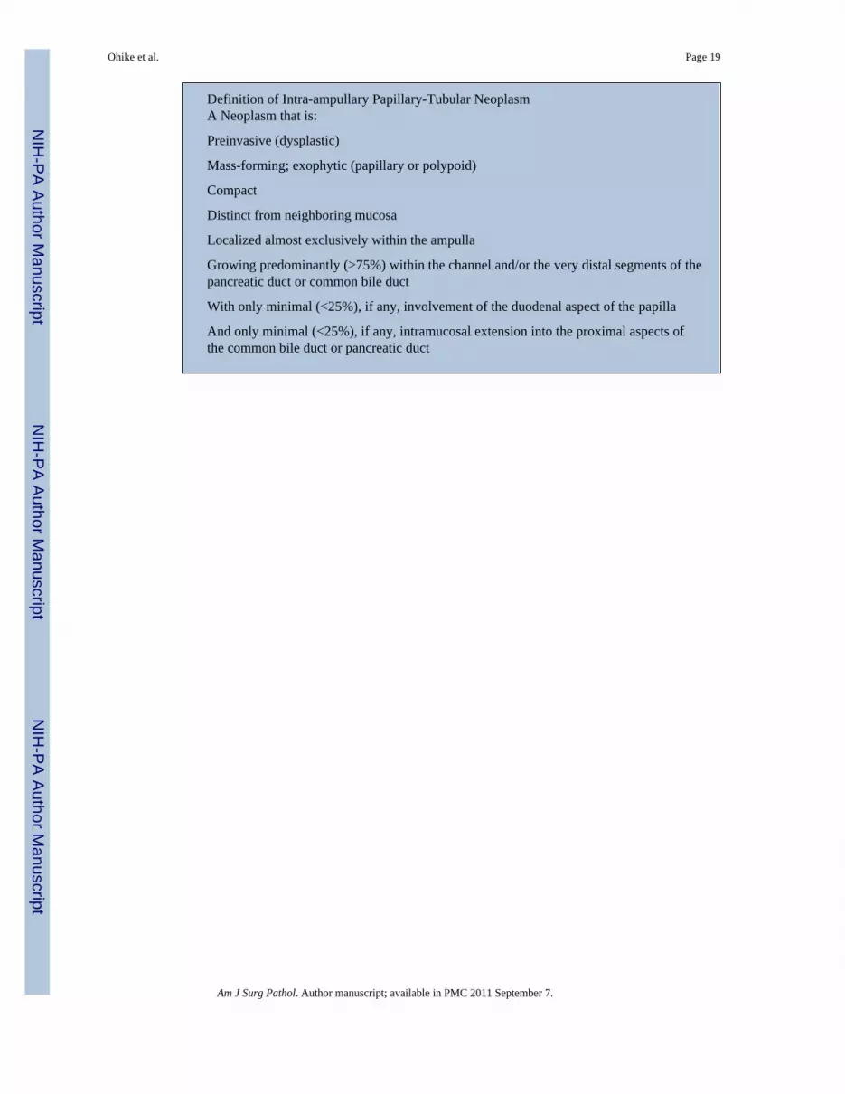
Definition of Intra-ampullary Papillary-Tubular NeoplasmA Neoplasm that is:
Preinvasive (dysplastic)
Mass-forming; exophytic (papillary or polypoid)
Compact
Distinct from neighboring mucosa
Localized almost exclusively within the ampulla
Growing predominantly (>75%) within the channel and/or the very distal segments of thepancreatic duct or common bile duct
With only minimal (<25%), if any, involvement of the duodenal aspect of the papilla
And only minimal (<25%), if any, intramucosal extension into the proximal aspects ofthe common bile duct or pancreatic duct
Ohike et al. Page 19
Am J Surg Pathol. Author manuscript; available in PMC 2011 September 7.
NIH
-PA Author Manuscript
NIH
-PA Author Manuscript
NIH
-PA Author Manuscript
FIGURE 1.A, Prominent exophytic growth dilating and filling the intra-ampullary ducts and formingobstructive papillary/ polypoid mass. B, By definition, there is only minimal (<25%) tumorat the duodenal surface of the papilla. CBD indicates common bile duct.
Ohike et al. Page 20
Am J Surg Pathol. Author manuscript; available in PMC 2011 September 7.
NIH
-PA Author Manuscript
NIH
-PA Author Manuscript
NIH
-PA Author Manuscript
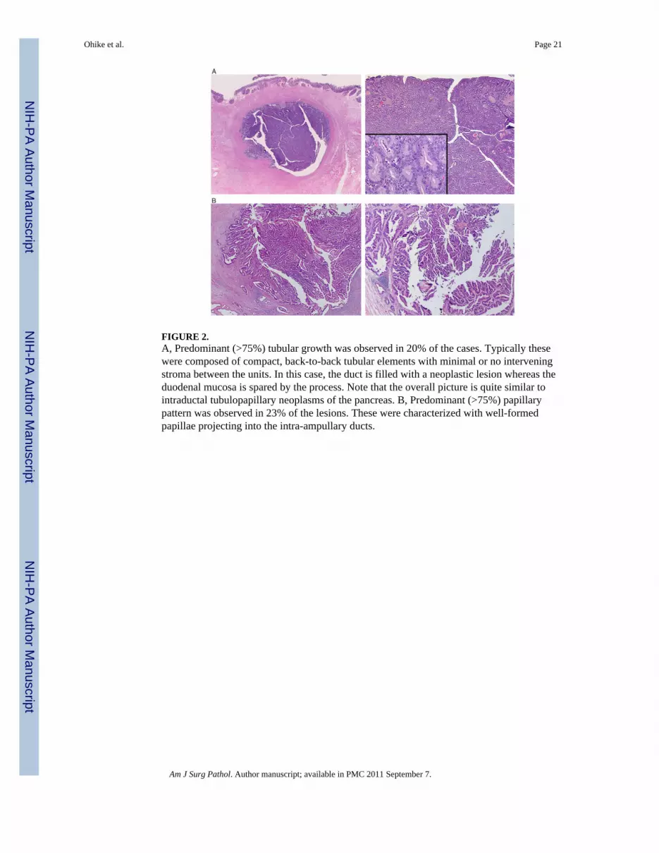
FIGURE 2.A, Predominant (>75%) tubular growth was observed in 20% of the cases. Typically thesewere composed of compact, back-to-back tubular elements with minimal or no interveningstroma between the units. In this case, the duct is filled with a neoplastic lesion whereas theduodenal mucosa is spared by the process. Note that the overall picture is quite similar tointraductal tubulopapillary neoplasms of the pancreas. B, Predominant (>75%) papillarypattern was observed in 23% of the lesions. These were characterized with well-formedpapillae projecting into the intra-ampullary ducts.
Ohike et al. Page 21
Am J Surg Pathol. Author manuscript; available in PMC 2011 September 7.
NIH
-PA Author Manuscript
NIH
-PA Author Manuscript
NIH
-PA Author Manuscript
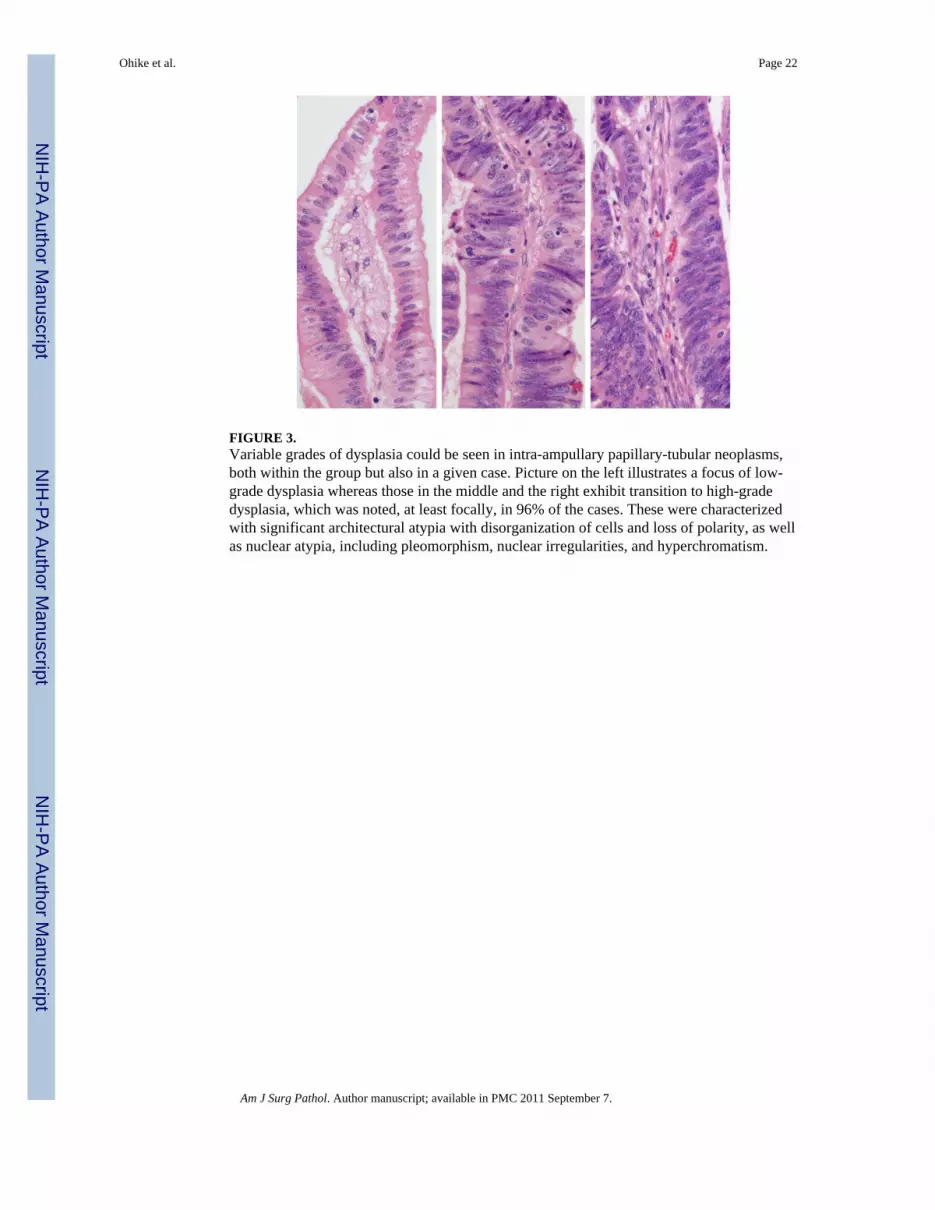
FIGURE 3.Variable grades of dysplasia could be seen in intra-ampullary papillary-tubular neoplasms,both within the group but also in a given case. Picture on the left illustrates a focus of low-grade dysplasia whereas those in the middle and the right exhibit transition to high-gradedysplasia, which was noted, at least focally, in 96% of the cases. These were characterizedwith significant architectural atypia with disorganization of cells and loss of polarity, as wellas nuclear atypia, including pleomorphism, nuclear irregularities, and hyperchromatism.
Ohike et al. Page 22
Am J Surg Pathol. Author manuscript; available in PMC 2011 September 7.
NIH
-PA Author Manuscript
NIH
-PA Author Manuscript
NIH
-PA Author Manuscript
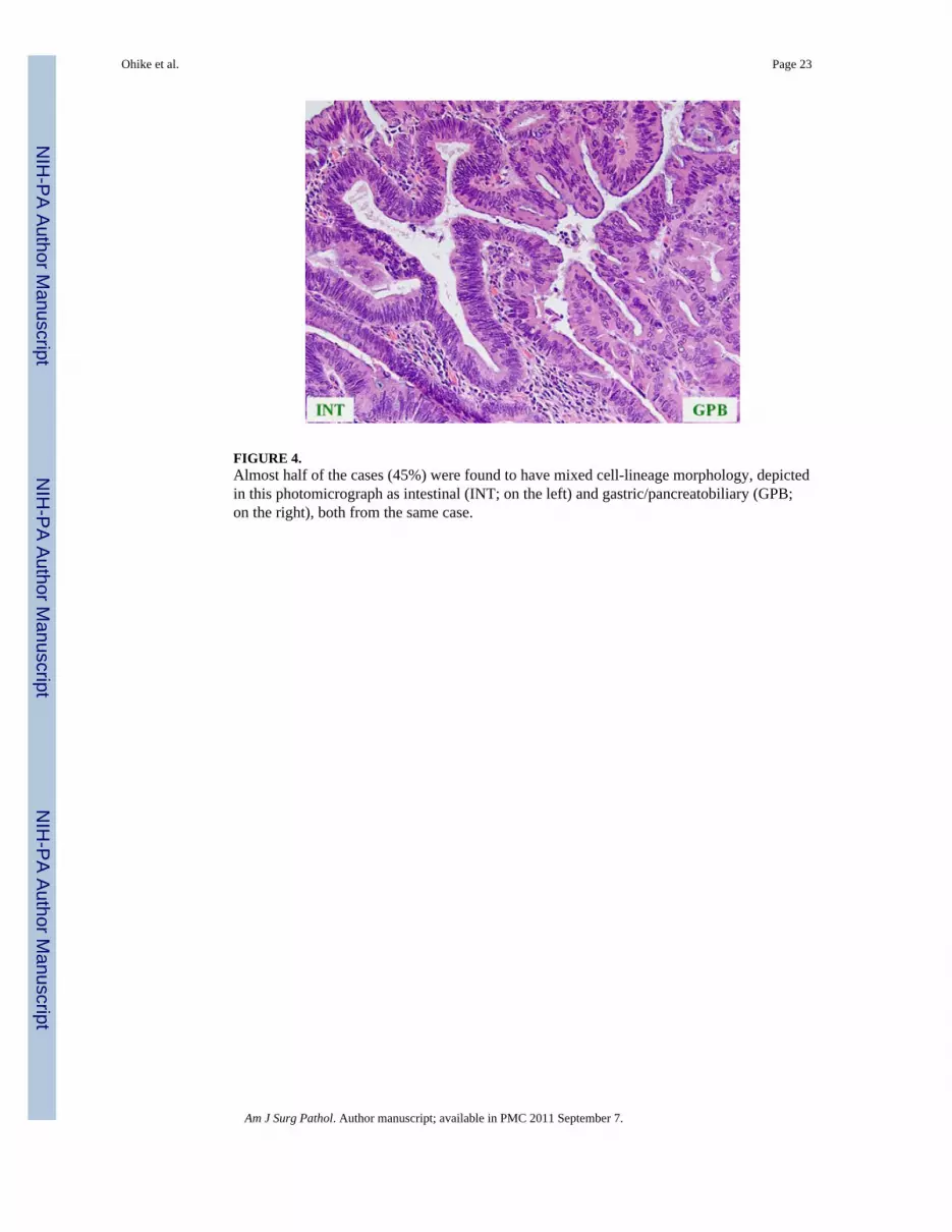
FIGURE 4.Almost half of the cases (45%) were found to have mixed cell-lineage morphology, depictedin this photomicrograph as intestinal (INT; on the left) and gastric/pancreatobiliary (GPB;on the right), both from the same case.
Ohike et al. Page 23
Am J Surg Pathol. Author manuscript; available in PMC 2011 September 7.
NIH
-PA Author Manuscript
NIH
-PA Author Manuscript
NIH
-PA Author Manuscript
FIGURE 5.A, Intestinal-type intra-ampullary papillary-tubular neoplasms were similar to conventionalcolonic/duodenal tubular adenomas consisting of relatively simple villous or tubularglandular units, lined by tall columnar cells with pseudostratified cigar-shaped nuclei. B,Goblet cells were prominent in some cases, and endocrine-type cells with distinctivegranules could also be seen.
Ohike et al. Page 24
Am J Surg Pathol. Author manuscript; available in PMC 2011 September 7.
NIH
-PA Author Manuscript
NIH
-PA Author Manuscript
NIH
-PA Author Manuscript
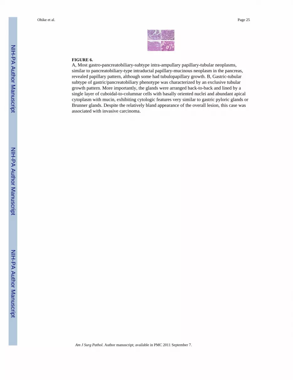
FIGURE 6.A, Most gastro-pancreatobiliary-subtype intra-ampullary papillary-tubular neoplasms,similar to pancreatobiliary-type intraductal papillary-mucinous neoplasm in the pancreas,revealed papillary pattern, although some had tubulopapillary growth. B, Gastric-tubularsubtype of gastric/pancreatobiliary phenotype was characterized by an exclusive tubulargrowth pattern. More importantly, the glands were arranged back-to-back and lined by asingle layer of cuboidal-to-columnar cells with basally oriented nuclei and abundant apicalcytoplasm with mucin, exhibiting cytologic features very similar to gastric pyloric glands orBrunner glands. Despite the relatively bland appearance of the overall lesion, this case wasassociated with invasive carcinoma.
Ohike et al. Page 25
Am J Surg Pathol. Author manuscript; available in PMC 2011 September 7.
NIH
-PA Author Manuscript
NIH
-PA Author Manuscript
NIH
-PA Author Manuscript
FIGURE 7.Invasive carcinomas were generally of tubular-type and were subclassified as pure intestinal(8%; left), pure pancreatobiliary type (20%; right), or mixed (72%; middle). Most cases with“mixed” features were classifiable as intestinal when a forced-binary (favored) approachwas used.
Ohike et al. Page 26
Am J Surg Pathol. Author manuscript; available in PMC 2011 September 7.
NIH
-PA Author Manuscript
NIH
-PA Author Manuscript
NIH
-PA Author Manuscript
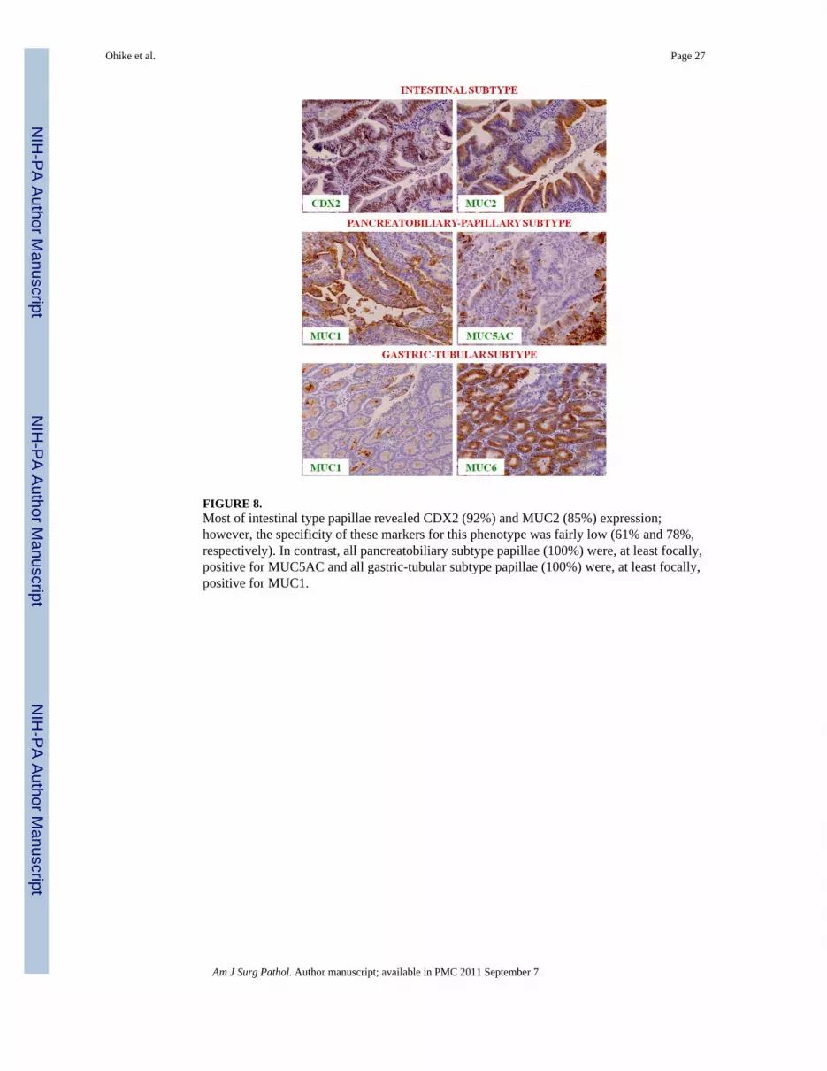
FIGURE 8.Most of intestinal type papillae revealed CDX2 (92%) and MUC2 (85%) expression;however, the specificity of these markers for this phenotype was fairly low (61% and 78%,respectively). In contrast, all pancreatobiliary subtype papillae (100%) were, at least focally,positive for MUC5AC and all gastric-tubular subtype papillae (100%) were, at least focally,positive for MUC1.
Ohike et al. Page 27
Am J Surg Pathol. Author manuscript; available in PMC 2011 September 7.
Am J Surg Pathol. Author manuscript; available in PMC 2011 September 7.
NIH
-PA Author Manuscript
NIH
-PA Author Manuscript
NIH
-PA Author Manuscript
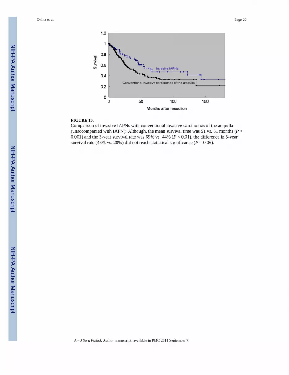
FIGURE 10.Comparison of invasive IAPNs with conventional invasive carcinomas of the ampulla(unaccompanied with IAPN): Although, the mean survival time was 51 vs. 31 months (P <0.001) and the 3-year survival rate was 69% vs. 44% (P < 0.01), the difference in 5-yearsurvival rate (45% vs. 28%) did not reach statistical significance (P = 0.06).
Ohike et al. Page 29
Am J Surg Pathol. Author manuscript; available in PMC 2011 September 7.
NIH
-PA Author Manuscript
NIH
-PA Author Manuscript
NIH
-PA Author Manuscript
NIH
-PA Author Manuscript
NIH
-PA Author Manuscript
NIH
-PA Author Manuscript
Ohike et al. Page 30
TABLE 1
Summary of Immunohistochemical Antibodies
Antigen Dilution Retrieval Source
MUC1 1:160 Citrate Leica, Microsystems; Bannockburn, IL
MUC2 1:100 Citrate Leica, Microsystems; Bannockburn, IL
MUC5AC 1:200 Trilog Leica, Microsystems; Bannockburn, IL
MUC6 1:80 Trilog Leica, Microsystems; Bannockburn, IL
CDX2 1:200 Citrate Biogenex, San Ramon, CA
CK7 1:40 Citrate DAKO, Carpintera, CA
CK20 1:40 Citrate DAKO, Carpintera, CA
CK indicates cytokeratin.
Am J Surg Pathol. Author manuscript; available in PMC 2011 September 7.
NIH
-PA Author Manuscript
NIH
-PA Author Manuscript
NIH
-PA Author Manuscript
Ohike et al. Page 31
TABLE 2
General Characteristics of the Cases With and Without Invasive Component