Risk of cancer in cases of suspected Lynch syndrome without germline mutation. SHORT TITLE: Cancer risk in suspected Lynch syndrome. AUTHORS: María Rodríguez-Soler (1-2), Lucía Pérez-Carbonell (2), Carla Guarinos (2), Pedro Zapater (3), Adela Castillejo (4), Victor M. Barberá (4), Miriam Juárez (2), Xavier Bessa (5), Rosa M. Xicola (6), Juan Clofent (7), Luis Bujanda (8), Francesc Balaguer (9), Josep-Maria Reñé (10), Luisa de-Castro (11), José C. Marín-Gabriel (12), Angel Lanas (13), Joaquín Cubiella (14), David Nicolás-Pérez (15), Alejandro Brea- Fernández (16), Sergi Castellví-Bel (9), Cristina Alenda (17), Clara Ruiz-Ponte (16), Angel Carracedo (16), Antoni Castells (9), Montserrat Andreu (5), Xavier Llor (6), José L. Soto (4), Artemio Payá (17), Rodrigo Jover (1). AUTHOR’S AFFILIATIONS 1. Unidad de Gastroenterología. Hospital General Universitario de Alicante. Alicante. Spain. 2. Unidad de Investigación. Hospital General Universitario de Alicante. Alicante. Spain. 3. Clinical Pharmacology Department. Hospital General Universitario de Alicante. Alicante. Spain. 4. Molecular Genetics Lab. Hospital General Universitario de Elche. Elche. Spain. 5. Gastroenterology Department. Hospital del Mar. Barcelona. Spain. 6. Department of Medicine and Cancer Center. University of Illinois at Chicago. Chicago, IL.
Transcript
Risk of cancer in cases of suspected Lynch syndrome without germline
mutation.
SHORT TITLE: Cancer risk in suspected Lynch syndrome.
AUTHORS: María Rodríguez-Soler (1-2), Lucía Pérez-Carbonell (2), Carla Guarinos
(2), Pedro Zapater (3), Adela Castillejo (4), Victor M. Barberá (4), Miriam Juárez (2),
Xavier Bessa (5), Rosa M. Xicola (6), Juan Clofent (7), Luis Bujanda (8), Francesc
Balaguer (9), Josep-Maria Reñé (10), Luisa de-Castro (11), José C. Marín-Gabriel (12),
Angel Lanas (13), Joaquín Cubiella (14), David Nicolás-Pérez (15), Alejandro Brea-
Carracedo, Castells, Andreu, Llor, Soto, and Payá.
Analysis and interpretation of data: Rodríguez-Soler, Zapater, Soto, Payá, and Jover.
Drafting of the manuscript: Rodríguez-Soler, Zapater, and Jover.
Critical revision of the manuscript for important intellectual content: Rodríguez-Soler,
Ruiz-Ponte, Carracedo, Castells, Andreu, Llor, Soto, and Jover.
Statistical analysis: Rodríguez-Soler, Zapater, and Jover.
5
ABSTRACT
Background & Aims: Colorectal cancers (CRC) with microsatellite instability (MSI)
and a mismatch repair (MMR) deficit without hypermethylation of the MLH1 promoter
are likely to be caused by Lynch syndrome. Some patients with these cancers have not
been found to have pathogenic germline mutations, and are considered to have Lynch-
like syndrome (LLS). The aim of this study was to determine the risk of cancer in
families of patients with LLS.
Methods: We studied a population-based cohort of 1705 consecutive patients,
performing MSI tests and immunohistochemical analyses of MMR proteins. Patients
were diagnosed with Lynch syndrome when they were found to have pathogenic
germline mutations. Patients with MSI and loss of MSH2 and/or MSH6 expression,
isolated loss of PMS2, or loss of MLH1 without MLH1 promoter hypermethylation and
no pathogenic mutation were considered to have LLS. The clinical characteristics of
patients and the age- and sex-adjusted standardized incidence ratios (SIR) of cancer in
families were compared between groups.
Results: The incidence of CRC was significantly lower in families of LLS patients than
families with confirmed Lynch syndrome cases (SIR for Lynch syndrome=6.04; 95%
confidence interval [CI], 3.58−9.54 and SIR for LLS=2.12; 95% CI, 1.16−3.56; P<.001).
However, the incidence of CRC was higher in families of patients with LLS than in
families with sporadic CRC (SIR for sporadic CRC=0.48; 95% CI, 0.27−0.79; P<.001).
Conclusions: The risk of cancer in families with LLS is lower that of families with
Lynch syndrome, but higher than that of families with sporadic CRC. These results
confirm the need for special screening and surveillance strategies for these patients
and their relatives.
6
Keywords : inherited colon cancer; cancer risk; genetic testing; immunohistochemistry
7
INTRODUCTION
Lynch Syndrome (LS) is the most common inherited colon cancer susceptibility
syndrome caused by germline mutations in one of several DNA mismatch repair (MMR)
genes, mainly MLH1 and MSH2, but also MSH6 and PMS2.1-3 Patients with LS have an
increased risk of colorectal cancer (CRC), endometrial cancer, and several other
cancers, including ovarian, upper urinary tract, gastric, small bowel, biliary/pancreatic,
skin, and brain cancers. The molecular signature of LS is microsatellite instability (MSI),
which is found in more than 95% of LS-associated CRCs.4 However, MSI is also
present in up to 15% of sporadic CRCs, due to hypermethylation of the promoter region
of MLH1 in tumor cells. Immunohistochemical (IHC) studies of MMR proteins have
been shown to be equivalent to MSI in detecting MMR-defective CRC.5 CRC with MSI
and a lack of staining of MSH2, MSH6, or MLH1 without promoter hypermethylation is
a strong indicator of MSH2, MSH6, or MLH1 germline mutations.6 However, some of
these CRC cases do not have pathogenic mutations in MMR genes. These cases are
suspected to be non-sporadic because no mechanism of inactivation is known for
these genes other than germline mutations in the context of LS. These patients are
considered to have “probably non-sporadic” colorectal cancer or “Lynch-like” syndrome
(LLS), and decisions about their management are not simple because of unconfirmed
suspicions of hereditary cancer. These patients must be distinguished from familial
CRC type X, where tumors do not show MMR deficiency. No studies have
characterized these CRC patients, and the risk of cancer in this group of families is not
known. Therefore, the surveillance strategy for these patients and their relatives is
unclear.
8
We analyzed the clinical and familial characteristics of patients diagnosed with LLS, LS,
or sporadic CRC. The main aim of this study was to determine the risk of cancer in
families of patients with LLS.
9
METHODS
Patients and data collection
The present study was a population-based observational cohort study including 1,705
patients with CRC from two nationwide multicenter studies, EPICOLON I and
EPICOLON II. EPICOLON I included consecutive patients with a new diagnosis of
CRC between November 2000 and October 2001 with the main goal of estimating the
incidence of LS in Spain.7 EPICOLON II also included consecutive newly diagnosed
CRC patients between March 2006 and December 2007 with the aim of investigating
different aspects related to the diagnosis of hereditary CRC.8 All of the patients
provided written informed consent. Both studies were approved by the institutional
review boards of the participating hospitals.
Patients were divided into three groups based on genetic data: 1) LS group, patients
with a confirmed pathogenic mutation in MLH1, MSH2, MSH6, or PMS2; 2) LLS group,
patients with MSI and loss of MSH2/MSH6 expression, isolated loss of PMS2, or loss
of expression of MLH1 without MLH1 promoter hypermethylation in which no germline
mutation was found; and 3) sporadic group, patients with CRC and MSS tumors
showing normal expression of MMR genes or a loss of MLH1 expression with MLH1
promoter hypermethylation.
Demographic, clinical, and pathology data were collected at the time of diagnosis.
Cancer pedigrees were built at diagnosis for CRC cases in the EPICOLON I and II
studies. The pedigrees were traced backward and laterally as far as possible. This
information was verified by reviewing medical records when available. Standardized
incidence ratios (SIR) for cancer were calculated as the ratio of the observed to
expected number of cases diagnosed in the families at the time of inclusion in the
EPICOLON I or II cohorts. In order to avoid recall bias, only cancer cases found in first-
10
degree relatives were included in the calculation of SIR. We considered tumors in the
endometrium, ovaries, upper urinary tract, stomach, small intestine, and hepatobiliary
system as non-colorectal LS-related cancers (LSRC). The index case was excluded for
the analysis of family history at the time of diagnosis. Calculation of the SIR was only
possible in families with complete pedigrees and information about the ages of all
family members, including relatives without cancer.
In 2011, the pedigrees were updated by asking patients and/or relatives about new
cancer cases after diagnosis of the index case. We include the index case for this
analysis, and the appearance of metachronous CRC or a new case of LSRC in the
index case was considered a new case in the family.
Microsatellite instability, immunohistochemical sta ining, and detection of
germline mutations
MSI analysis was performed in all patients. We ascertained MSI status using BAT26
and NR24 quasimonomorphic markers as described previously.9 MSI was present
when one of the two markers was unstable. IHC analysis of the four mismatch repair
proteins MLH1, MSH2, MSH6, and PMS2 in tumor tissue was performed in all patients
using tissue microarrays (TMAs) as described previously.10 In patients with a loss of
MLH1, methylation of MLH1 and BRAF mutation status were analyzed. MLH1
methylation analysis was performed using methylation-specific multiplex ligation-
dependent probe amplification (MS-MLPA) according to the manufacturer´s protocol
using the SALSA MS-MLPA kit ME011 Mismatch Repair Genes (MRC-Holland,
Amsterdam, the Netherlands).11 The V600E BRAF mutation was detected using
specific TaqMan probes in real-time PCR (ABI PRISM 7500, Applied Biosystems,
Foster City, CA, USA) and allelic discrimination software as described previously.12
11
Germline mutation analysis was performed in accordance with the results of the IHC
analysis as described previously.10 Patients with loss of MSH2 expression with no
detected mutation were analyzed for EPCAM rearrangements using MLPA (MRC-
Holland, Amsterdam, the Netherlands) according to the manufacturer's recommended
protocol. DNA sequencing was performed to characterize the deletion breakpoints.13
Large rearrangements (deletions and insertions) were tested using MLPA (MRC-
Holland) according to the manufacturer´s protocol. The genetic analysis results were
interpreted based on the ACMG Recommendations for Standards for Interpretation of
Sequence Variations (2000) and the InSIGHT database.14
Statistical analysis
Continuous variables are reported as mean ± standard deviation (SD) or medians and
25th and 75th percentiles for non-normally distributed data. Categorical variables are
reported as frequencies or percentages. Significant differences between groups were
analyzed using the chi-square test for categorical data and the non-parametric Mann-
Whitney U test for quantitative data.
The SIR of each cancer was calculated as the ratio of the observed to expected
number of cases among relatives. Person-years were calculated from 20 years of age
until the earliest cancer diagnosis or death. The expected number of cases was
calculated as the sum of the products of the number of person-years for each 5-year
age/sex group and the corresponding age/sex-specific incidence rates in Spanish
regional registers.15 The confidence limits were based on the Byar's approximation of
the exact Poisson distribution, which is accurate even with small numbers.16 All
reported p-values are two-sided, and p < 0.05 was considered significant. All
calculations were performed using the SPSS 19.0 software.
12
RESULTS
A total of 1,705 patients with CRC were included in the study. The median age was 71
years (range: 27-101 years) and 59% of patients were male. Sixteen patients were
excluded because of discrepancies between the IHC and MSI analyses; no mutation
was found in these patients. Therefore, data from 1,689 patients were analyzed.
Tumors from all patients were analyzed. A total of 135 patients (8%) exhibited in
their tumors MSI and loss of expression of any of the MMR proteins. In 104 patients
(6.1%), loss of MLH1 expression was found in IHC analysis. Of these patients, 25
(1.4%) did not exhibit hypermethylation of the promoter region. We also used BRAF
mutation as a sporadic CRC marker in these 25 cases, but no case of BRAF mutation
indicating sporadic origin was found. Loss of MSH2 expression was seen in the IHC
analysis of 22 patients (1.3%). Three patients (0.2%) had an isolated loss of MSH2
expression, and 19 (1.1%) had a combined loss of MSH2 and MSH6. Isolated loss of
MSH6 was found in 6 (0.3%) patients. Finally, an isolated loss of PMS2 was found in 3
patients (0.2%).
A germline pathogenic mutation in any of the MMR genes was found in 16 patients
(0.9%) and considered to have LS. Three of these patients exhibited MSI with non-
valuable expression of MMR proteins in IHC analysis. Four of the LS patients were
found to have a pathogenic mutation in MLH1, eight in MSH2, three in MSH6, and one
in PMS2. All of these patients exhibited MSI. No case was found with deletions in
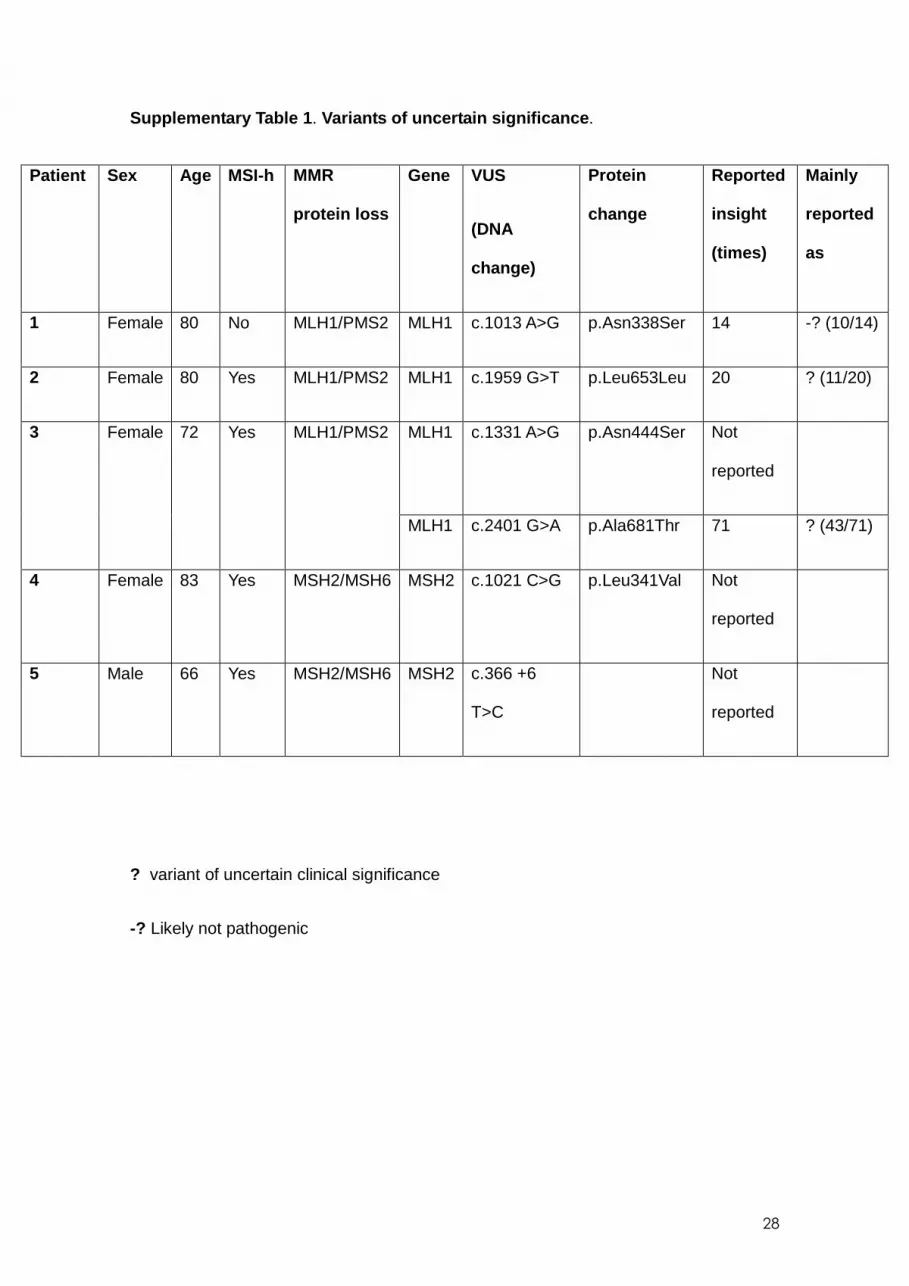
EPCAM. Variants of uncertain significance were found in five patients (Supplementary
Table 1) . Forty-three patients (2.5%) exhibited MSI and loss of MSH2/MSH6, PMS2, or
MLH1 expression without promoter hypermethylation, but no pathogenic germline
mutation was found. These patients were considered to have LLS. Among the LLS
patients, 21 were found to have a loss of MLH1 protein expression, and 22 loss of
13
MSH2, MSH6, or PMS2 expression (14 with loss of MSH2 and MSH6, 6 with isolated
loss of MSH6, and 2 with isolated loss of PMS2). Finally, 1,630 patients (96%) were
considered to have sporadic CRC.
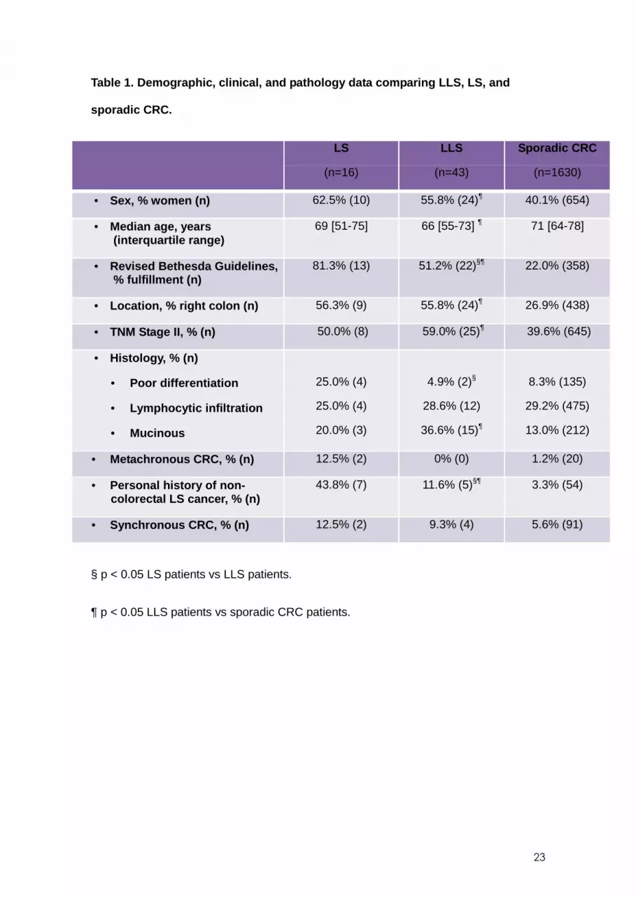
Demographic, clinical, and pathological characteris tics of patients with LLS
The characteristics of the LLS group (n=43) were compared to the LS (n=16) and
sporadic CRC (n=1630) groups (Table 1 ). Fewer LLS patients fulfilled the revised
Bethesda guidelines than LS patients, and LLS patients less often had a personal
history of non-colorectal LSRC compared to LS patients. No differences were found in
the presence of metachronous CRC, median age at diagnosis of CRC, sex and tumor
characteristics, such as location or TNM classification. When we compared LLS
patients to sporadic CRC patients, patients with LLS were younger at diagnosis,
predominantly female, and more frequently fulfilled the revised Bethesda guidelines.
Personal history of non-colorectal LSRC was more frequent in patients with LLS
without differences in the presence of metachronous or synchronous CRC (Table 1 ).
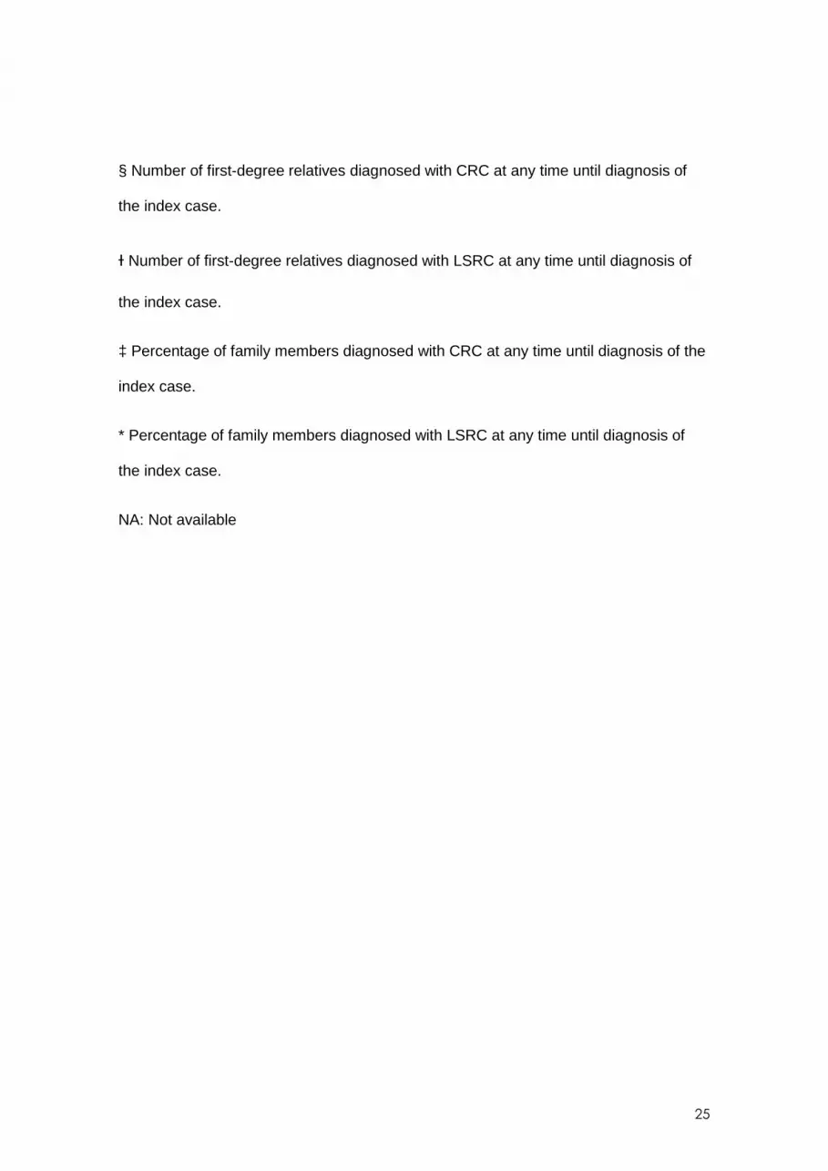
Familial cancer risk
A total of 13 families with LS and 25 families with LLS had complete pedigrees
including the ages of relatives without cancer. A random sample of 115 families with
sporadic CRC was used for a comparison. A total of 1,102 first-degree relatives were
included: 80 from LS families, 177 from LLS families, and 845 from sporadic CRC
families. The mean number of first-degree relatives was 6.1 for LS families, 7.0 in LLS
families, and 7.3 in sporadic CRC families.
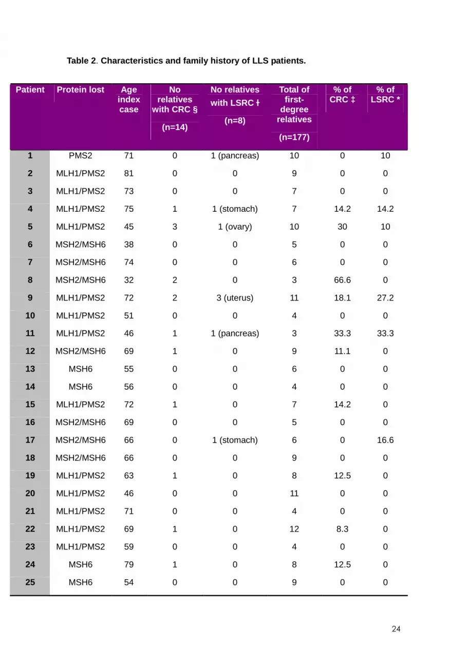
In LS families we identified 18 cases of CRC and six cases of non-colorectal LSRC.
Only in 4 (30.7%) families there was no cancer case other than the index case. In LLS
14
families, we found 14 cases of CRC and eight cases of non-colorectal LSRC. In 12 out
of 25 families (48%), no case of cancer was found other than the index case. The
characteristics of patients and distribution of cancer cases in LLS families are provided
in Table 2 . Finally, in sporadic CRC families, 15 first-degree relatives had CRC and 27
had non-colorectal LSRC. In 85 (79.9%) families no cancer cases were identified other
than the index case.
The SIR of CRC and non-colorectal LSRC are found in Table 3 . The incidence of CRC
was significantly lower in LLS families compared to confirmed LS families (SIR in LS:
6.04, 95% CI 3.58-9.54; SIR in LLS: 2.12, 95% CI 1.16-3.56; p<0.001). However, the
incidence of CRC was significantly greater in the LLS families than in sporadic CRC
families (SIR in sporadic CRC: 0.48, 95% CI 0.27-0.79; p<0.001). The SIR for non-
colorectal LSRC was non-significantly higher in LS families (SIR 2.81, 95% CI 1.03-
6.12) compared to LLS families (SIR 1.69, 95% CI 0.73-3.34; p=0.09). There were no
differences in SIR for non-colorectal LSRC between LLS families and sporadic
CRC families (SIR 1.20, 95% CI 0.79-1.74; p=0.5). Taken together, the results indicate
that, for CRC and non-colorectal LSRC, the highest risk is for LS families (SIR 4.69;
95% CI 3.00-6.98), followed by LLS families (SIR 1.94; 95% CI 1.22-2.94; p<0.001).
The risk in LLS families was significantly higher than the risk in relatives of patients with
sporadic CRC (SIR 0.78; 95% CI 0.56-1.05; p<0.001).
Figure 1 shows the cumulative age-of-onset curves for CRC among all relatives in the
LS, LLS, and sporadic CRC groups. The relatives of patients with LLS developed CRC
at an earlier mean age (53.71±16.8 years) than those with sporadic CRC (68.8±9
years; p=0.004), but was similar to the mean age in LS patients (48.5±14.13; p=0.23).
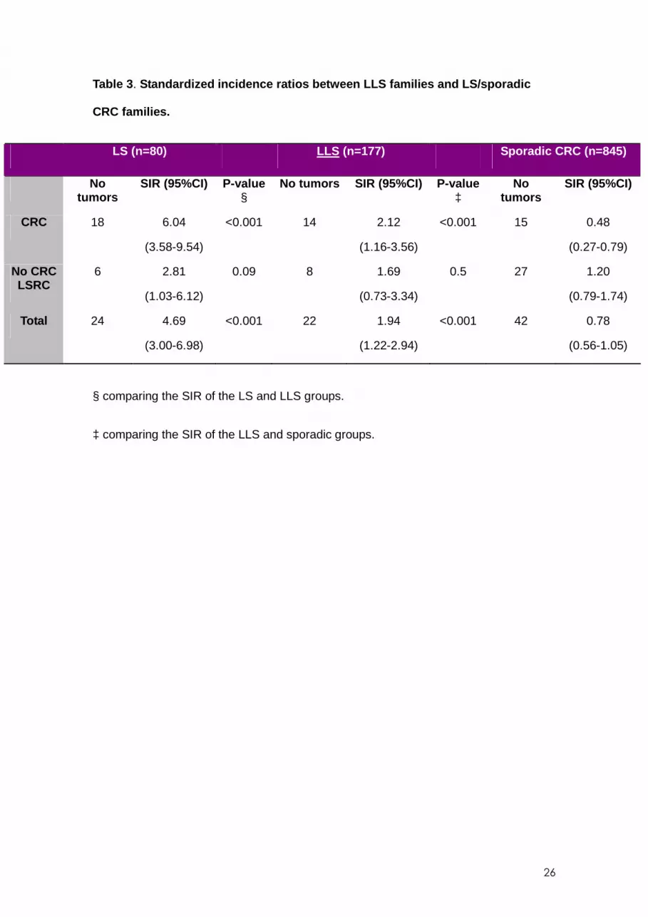
After a median 8.3 years of prospective follow-up, cancer pedigrees were updated in
93 families: 10 in the LS group, 16 in the LLS group, and 67 in the sporadic CRC. A
total of 533 first-degree relatives were included (including the index case): 41 from LS
15
families, 89 from LLS families, and 403 from sporadic CRC families. During this period,
7 (17.1%) new cases of CRC or non-colorectal LSRC appeared in LS families, 4 (4.5%)
new cases in LLS families, and 4 (1.0%) in families with sporadic CRC (Table 4).
16
DISCUSSION
The main finding of our study is that the risk of CRC is lower in LLS families than
among patients with genetically confirmed LS but significantly higher than in cases of
sporadic CRC. The results confirm the need for a special surveillance strategy for these
patients and their relatives. In addition, the age of onset for CRC in LLS families was
similar to that of LS families. Differences between LLS families and families with
sporadic CRC cases were more prominent in regards to CRC risk than for the risk of
other LSRCs.
Recent studies have shown that MSI testing and IHC analysis of MMR genes in
all patients with CRC improves the detection of patients with LS.10, 17, 18 Because of the
generalization of this universal strategy following Jerusalem guidelines,19 an increasing
number of patients exhibit a loss of MMR protein expression with no pathogenic
mutation. In CRC cases with a loss of MSH2, MSH6, PMS2, or MLH1 without
hypermethylation, no cause of MMR gene inactivation is known other than germline
mutation. In these cases, when a germline mutation is not found, a high suspicion of LS
persists, and these patients and their relatives should be followed-up appropriately. The
clinical characteristics of some of these patients suggest that they are clear hereditary
cases, even though we were not able to find a genetic defect. Some of the pedigrees of
patients with LLS showed a significant history of CRC with metachronous and
synchronous tumors and fulfillment of the Amsterdam criteria. In these cases, the
genetic defect was not found, probably due to it being located in a still unknown part of
the gene, or simply that our technical capacity is not yet able to detect the pathogenic
mutation. Some of these cases have been explained in the literature by alterations in
other genes, such as in cases of EPCAM deletions or MLH1 constitutional
epimutations. Other mechanisms, including inversions and duplications, could also
explain some of these cases.20-26 However, some cases do not show any specific
characteristics that suggest a hereditary origin. A notable proportion of LLS families do
17
not have a history of other cancers, and the only reason to suspect LS is the presence
of MSI and loss of MMR protein expression. In these cases, determining the
appropriate counseling for patients and their relatives is difficult. It is possible that
some of these LLS patients could be cases with CRC who may have false
positive results of IHC and/or MSI or sporadic MMR- deficient CRC, and LLS
patients would be a mixture of true LS patients wit h non-detected germline
mutation and sporadic CRC cases. However , the high risk of CRC found in our study
suggests that, in its entirety, LLS cases should be considered as high-risk cases and
strategies for cancer prevention must be implemented in this group of patients and their
relatives. The SIR of CRC for LLS families was similar to that described in a group of
families with familial colorectal cancer syndrome type X, but in this syndrome no
molecular alteration has been found.27 Even though LLS is a completely different entity
because of the presence of MSI, the similar risk of CRC cancer should guarantee at
least a similar surveillance program, even in cases without previous family history. Our
results can contribute some rationale for designing follow-up strategies and, together
with family history, can help clinicians appropriately schedule surveillance for these
patients and their relatives. In our study, the age of CRC onset was similar to that of
LS, and therefore surveillance strategies should start at the same age as
recommended for LS cases. On the other hand, the frequency of CRC screening
should be individualized. Given that the risk of CRC is lower in LLS families than in LS
families, longer surveillance intervals for LLS cases and relatives without a prominent
family history of CRC may be recommended. We have not found higher risk for non-
colorectal LSRC in LLS families compared to sporadi c CRC families. However,
that can be due to the small number of cases detect ed in our series. For this
reason, specific recommendations for endometrial an d other non-colorectal
LSRC cannot be appropriately supported by our data.
18
The limitations of our study are the possibility of underreporting or misreporting cancers
because our information was not always confirmed with objective clinical and
pathological data. However, we think this limitation is minor because it would affect the
LLS group to the same extent as the other groups. Another limitation is the relatively
small number of families, especially in the prospectively followed cases, which
precludes finding clear differences between groups. Moreover, the follow-up time for
these cases could be considered too short.
The main strength of our study is its population-based approach with cases ascertained
from general clinics and not from specialized high-risk clinics. This approach provides
robustness to our data in terms of potential applicability to general practice. Cancer risk
can be overestimated in studies coming from select populations in genetic high-risk
clinics. Studies based on recruitment through cancer genetics clinics do not usually
correct for the selection bias caused by the over-representation of families with multiple
cases in the data set.28, 29 Our results attempt to provide a rationale for follow-up and
surveillance of this growing group of patients that will mostly be seen in the general
clinics and not in high-risk clinics. New research is necessary to refine the classification
of these patients in order to distinguish between sporadic and true hereditary cases.
19
Figure 1. Cumulative age of onset of colorectal cancer in first-degree relatives of Lynch
syndrome, Lynch-like syndrome, and sporadic CRC.
20
Reference List
1. Aarnio M, Sankila R, Pukkala E, et al. Cancer risk in mutation carriers of DNA-mismatch-repair genes. Int J Cancer 1999;81:214-218.
2. Bonadona V, Bonaiti B, Olschwang S, et al. Cancer risks associated with germline mutations in MLH1, MSH2, and MSH6 genes in Lynch syndrome. JAMA 2011;305:2304-2310.
3. Watson P, Lynch HT. Extracolonic cancer in hereditary nonpolyposis colorectal cancer. Cancer 1993;71:677-685.
4. Aaltonen LA, Peltomaki P, Mecklin JP, et al. Replication errors in benign and malignant tumors from hereditary nonpolyposis colorectal cancer patients. Cancer Res 1994;54:1645-1648.
5. Pinol V, Castells A, Andreu M, et al. Accuracy of revised Bethesda guidelines, microsatellite instability, and immunohistochemistry for the identification of patients with hereditary nonpolyposis colorectal cancer. JAMA 2005;293:1986-1994.
6. Lynch HT, Boland CR, Rodriguez-Bigas MA, et al. Who should be sent for genetic testing in hereditary colorectal cancer syndromes? J Clin Oncol 2007;25:3534-3542.
7. Pinol V, Andreu M, Castells A, et al. Frequency of hereditary non-polyposis colorectal cancer and other colorectal cancer familial forms in Spain: a multicentre, prospective, nationwide study. Eur J Gastroenterol Hepatol 2004;16:39-45.
8. Abuli A, Bessa X, Gonzalez JR, et al. Susceptibility genetic variants associated with colorectal cancer risk correlate with cancer phenotype. Gastroenterology 2010;139:788-96, 796.
9. Xicola RM, Llor X, Pons E, et al. Performance of different microsatellite marker panels for detection of mismatch repair-deficient colorectal tumors. J Natl Cancer Inst 2007;99:244-252.
10. Perez-Carbonell L, Ruiz-Ponte C, Guarinos C, et al. Comparison between universal molecular screening for Lynch syndrome and revised Bethesda guidelines in a large population-based cohort of patients with colorectal cancer. Gut 2012;61:865-872.
11. Perez-Carbonell L, Alenda C, Paya A, et al. Methylation analysis of MLH1 improves the selection of patients for genetic testing in Lynch syndrome. J Mol Diagn 2010;12:498-504.
12. Benlloch S, Paya A, Alenda C, et al. Detection of BRAF V600E mutation in colorectal cancer: comparison of automatic sequencing and real-time chemistry methodology. J Mol Diagn 2006;8:540-543.
13. Van der KH, Wijnen J, Wagner A, et al. Molecular characterization of the spectrum of genomic deletions in the mismatch repair genes MSH2, MLH1,
21
MSH6, and PMS2 responsible for hereditary nonpolyposis colorectal cancer (HNPCC). Genes Chromosomes Cancer 2005;44:123-138.
14. Ou J, Niessen RC, Vonk J, et al. A database to support the interpretation of human mismatch repair gene variants. Hum Mutat 2008;29:1337-1341.
15. Chirlaque-Lopez MD, Salmeron-Martinez D, Valera-Ninirola I, et al. Incidencia de cancer en la Region de Murcia. In: Direccion General de Salud Publica.Consejeria de Sanidad de la Region de Murcia, ed. 2007.
16. Breslow NE, Day NE. Statistical methods in cancer research. IARC Workshop 25-27 May 1983. IARC Sci Publ 1987;1-406.
17. Hampel H, Frankel WL, Martin E, et al. Screening for the Lynch syndrome (hereditary nonpolyposis colorectal cancer). N Engl J Med 2005;352:1851-1860.
18. Hampel H, Frankel WL, Martin E, et al. Feasibility of screening for Lynch syndrome among patients with colorectal cancer. J Clin Oncol 2008;26:5783-5788.
19. Boland CR, Shike M. Report from the Jerusalem workshop on Lynch syndrome-hereditary nonpolyposis colorectal cancer. Gastroenterology 2010;138:2197.
20. Chen JM. The 10-Mb paracentric inversion of chromosome arm 2p in activating MSH2 and causing hereditary nonpolyposis colorectal cancer: re-annotation and mutational mechanisms. Genes Chromosomes Cancer 2008;47:543-545.
21. Guarinos C, Castillejo A, Barbera VM, et al. EPCAM germ line deletions as causes of Lynch syndrome in Spanish patients. J Mol Diagn 2010;12:765-770.
22. Kovacs ME, Papp J, Szentirmay Z, et al. Deletions removing the last exon of TACSTD1 constitute a distinct class of mutations predisposing to Lynch syndrome. Hum Mutat 2009;30:197-203.
23. Ligtenberg MJ, Kuiper RP, Chan TL, et al. Heritable somatic methylation and inactivation of MSH2 in families with Lynch syndrome due to deletion of the 3' exons of TACSTD1. Nat Genet 2009;41:112-117.
24. Morak M, Koehler U, Schackert HK, et al. Biallelic MLH1 SNP cDNA expression or constitutional promoter methylation can hide genomic rearrangements causing Lynch syndrome. J Med Genet 2011;48:513-519.
25. Pinheiro M, Pinto C, Peixoto A, et al. A novel exonic rearrangement affecting MLH1 and the contiguous LRRFIP2 is a founder mutation in Portuguese Lynch syndrome families. Genet Med 2011;13:895-902.
26. Wagner A, van der KH, Franken P, et al. A 10-Mb paracentric inversion of chromosome arm 2p inactivates MSH2 and is responsible for hereditary nonpolyposis colorectal cancer in a North-American kindred. Genes Chromosomes Cancer 2002;35:49-57.
27. Lindor NM, Rabe K, Petersen GM, et al. Lower cancer incidence in Amsterdam-I criteria families without mismatch repair deficiency: familial colorectal cancer type X. JAMA 2005;293:1979-1985.
28. Bonadona V, Bonaiti B, Olschwang S, et al. Cancer risks associated with germline mutations in MLH1, MSH2, and MSH6 genes in Lynch syndrome. JAMA 2011;305:2304-2310.
29. Carayol J, Khlat M, Maccario J, et al. Hereditary non-polyposis colorectal cancer: current risks of colorectal cancer largely overestimated. J Med Genet 2002;39:335-339.
23
Table 1. Demographic, clinical, and pathology data comparing LLS, LS, and
4 Female 83 Yes MSH2/MSH6 MSH2 c.1021 C>G p.Leu341Val Not
reported
5 Male 66 Yes MSH2/MSH6 MSH2 c.366 +6
T>C
Not
reported
29
ANNEX. Study participants: Members of the EPICOLON Consort ium (Gastrointestinal Oncology Group of the Spanish Gastroenterological Association). Hospital 12 de Octubre, Madrid: Juan Diego Morillas (local coordinator), Raquel Muñoz, Marisa Manzano, Francisco Colina, Jose Díaz, Carolina Ibarrola, Guadalupe López, Alberto Ibáñez; Hospital Clínic, Barcelona: Antoni Castells (local coordinator), Virgínia Piñol, Sergi Castellví-Bel, Francesc Balaguer, Victoria Gonzalo, Teresa Ocaña, María Dolores Giráldez, Maria Pellisé, Anna Serradesanferm, Leticia Moreira, Miriam Cuatrecasas, Josep M. Piqué; Hospital Clínico Universitario, Zaragoza: Ángel Lanas (local coordinator), Javier Alcedo, Javier Ortego; Hospital Cristal-Piñor, Complexo Hospitalario de Ourense: Joaquin Cubiella (local coordinator), Mª Soledad Díez, Mercedes Salgado, Eloy Sánchez, Mariano Vega; Hospital del Mar, Barcelona: Montserrat Andreu (local coordinator), Anna Abuli, Xavier Bessa, Mar Iglesias, Agustín Seoane, Felipe Bory, Gemma Navarro, Beatriz Bellosillo; Josep Mª Dedeu, Cristina Álvarez, Begoña Gonzalez; Hospital San Eloy, Baracaldo and Hospital Donostia, CIBERehd, University of Country Basque, San Sebastián: Luis Bujanda (local coordinator) Ángel Cosme, Inés Gil, Mikel Larzabal, Carlos Placer, María del Mar Ramírez, Elisabeth Hijona, Jose M. Enríquez-Navascués y Jose L. Elosegui; Hospital General Universitario de Alicante: Artemio payá (EPICOLON I local coordinator), Rodrigo Jover (EPICOLON II local coordinator), Cristina Alenda, Laura Sempere, Nuria Acame, Estefanía Rojas, Lucía Pérez-Carbonell; Hospital General de Granollers: Joaquim Rigau (local coordinator), Ángel Serrano, Anna Giménez; Hospital General de Vic: Joan Saló (local coordinator), Eduard Batiste-Alentorn, Josefina Autonell, Ramon Barniol; Hospital General Universitario de Guadalajara and Fundación para la Formación e Investigación Sanitarias Murcia: Ana María García (local coordinator), Fernando Carballo, Antonio Bienvenido, Eduardo Sanz, Fernando González, Jaime Sánchez, Akiko Ono; Hospital General Universitario de Valencia: Mercedes Latorre (local coordinator), Enrique Medina, Jaime Cuquerella, Pilar Canelles, Miguel Martorell, José Ángel García, Francisco Quiles, Elisa Orti; CHUVI-Hospital Meixoeiro, Vigo: EPICOLON I: Juan Clofent (local coordinator), Jaime Seoane, Antoni Tardío, Eugenia Sanchez. EPICOLON II Mª Luisa de Castro (local coordinator), Antoni Tardío, Juan Clofent, Vicent Hernández; Hospital Universitari Germans Trias i Pujol, Badalona and Section of Digestive Diseases and Nutrition, University of Illinois at Chicago, IL, USA. : Xavier Llor (local coordinator), Rosa M. Xicola, Marta Piñol, Mercè Rosinach, Anna Roca, Elisenda Pons, José M. Hernández, Miquel A. Gassull; Hospital Universitari Mútua de Terrassa: Fernando Fernández-Bañares (local coordinator), Josep M. Viver, Antonio Salas, Jorge Espinós, Montserrat Forné, Maria Esteve; Hospital Universitari Arnau de Vilanova, Lleida: Josep M. Reñé (local coordinator), Carmen Piñol, Juan Buenestado, Joan Viñas; Hospital Universitario de Canarias: Enrique Quintero (local coordinator), David Nicolás, Adolfo Parra, Antonio Martín; Hospital Universitario La Fe, Valencia: Lidia Argüello (local coordinator), Vicente Pons, Virginia Pertejo, Teresa Sala; Hospital Sant Pau, Barcelona: Dolors Gonzalez (local coordinator) Eva Roman, Teresa Ramon, Maria Poca, Mª Mar Concepción, Marta Martin, Lourdes Pétriz; Hospital Xeral Cies, Vigo: Daniel Martinez (local coordinator); Fundacion Publica Galega de Medicina Xenomica (FPGMX), CIBERER, Genomic Medicine Group-University of Santiago de Compostela, Santiago de Compostela, Galicia, Spain: Ángel Carracedo (local coordinator), Clara Ruiz-Ponte, Ceres Fernández-Rozadilla, Mª Magdalena Castro; Hospital Universitario Central de Asturias: Sabino Riestra (local coordinator), Luis Rodrigo; Hospital de Galdácano, Vizcaya: Javier Fernández (local
30
coordinator), Jose Luis Cabriada; Fundación Hospital de Calahorra (La Rioja) La Rioja: Luis Carreño (local coordinator), Susana Oquiñena, Federico Bolado; Hospital Royo Villanova, Zaragoza: Elena Peña (local coordinator), José Manuel Blas, Gloria Ceña, Juan José Sebastián; Hospital Universitario Reina Sofía, Córdoba: Antonio Naranjo (local coordinator).