Original Article Extra-virgin olive oil-enriched diet modulates DSS-colitis-associated colon carcinogenesis in mice S. Sánchez-Fidalgo, I. Villegas a , A. Cárdeno, E. Talero, M. Sánchez-Hidalgo, V. Motilva, C. Alarcón de la Lastra * Department of Pharmacology, Faculty of Pharmacy, University of Seville, Profesor García González Street, 2. 41012-Seville, Spain article info Article history: Received 16 June 2009 Accepted 3 March 2010 Keywords: Extra virgin olive oil Ulcerative colitis Colorectal cancer b-catenin Inflammation parameters summary Background & aims: Patients with inflammatory bowel disease (IBD) are at increased risk for developing ulcerative colitis (UC)-associated colorectal cancer (CRC). Several studies have shown that extra virgin olive oil (EVOO) might possess anti-inflammatory, antiproliferative and antiapoptotic effects. We have evaluated EVOO diet effects on the severity of repeated colitis-associated CRC. Methods: Six-week-old C57BL/6 mice were randomized into two dietary groups: sunflower oil (SFO) and EVOO diets, both at 10%. Mice were exposed to 15 cycles of 0.7% dextran sodium sulphate (DSS) for 1 week followed by distilled water for 10 days. After, the rats were sacrificed and colonic damage was both histologically and biochemically assessed. Results: Disease activity index (DAI) was significantly higher on SFO vs. EVOO diet at the end of the experimental period. EVOO-fed mice showed less incidence and multiplicity of tumors than in those SFO-fed mice. b-catenin immunostaining was limited to cell membranes in control groups, whereas translocation from the cell membrane to the cytoplasm and/or nucleus was showed in DSS-treated groups and its expression was higher in SFO-fed animals. Cytokine production was significantly enhanced in SFO-fed mice, while this increase was not significant in EVOO-fed mice. Conversely, cyclooxigenase-2 (COX-2) and inducible nitric oxidase synthase (iNOS) expression were significantly lower in the animal group fed with EVOO than in the SFO group. Conclusions: These results confirm that EVOO diet has protective/preventive effect in the UC-associated CRC. This beneficial effect was correlated with a better DAI, a minor number of dysplastic lesions, a lower b-catenin immunoreactivity, a proinflammatory cytokine levels reduction, a non modification of p53 expression and, COX-2 and iNOS reduction in the colonic tissue. Ó 2010 Elsevier Ltd and European Society for Clinical Nutrition and Metabolism. All rights reserved. 1. Introduction Colorectal cancer (CRC) is one of the most common gastro- intestinal tract malignancies. It is the third cause of cancer- related death in the Western world and affects about one million people every year throughout the world with a high mortality rate. CRC develops from a dysplastic precursor lesion, sporadi- cally, in the context of high-risk hereditary conditions, or in the background of chronic inflammation. In effect, patients with inflammatory bowel disease (IBD) are among the highest risk groups for developing CRC. The risk of colorectal malignancies in colitis patients increases with the extent and duration of the disease. 1,2 At present no genetic basis is able to explain the predisposition to CRC in those patients. Nevertheless, the main genomic instability that contribute to colon carcinogenesis is chromosomal instability, which results in damage of genetic material and consequently, loss of function of key tumor suppressor genes such as p53, which is expressed or expresses proteins that regulate growth and apoptosis. b-catenin is also an important cancer target, which plays a role in both cell adhesion and intracellular signalling. It is known to be a key component of the cadherin mediated-cell adhesion system and an important molecule in Wnt-APC signal transduction system. 3,4 Several lines of evidence implicate chronic inflammation as a key predisposing factor to CRC in IBD. The inducible isoforms of cyclooxygenase (COX-2) and nitric oxidase synthase (iNOS) are the main enzymes involved. COX-2 is expressed after proinflammatory cytokines stimulation, growth factors, reactive oxygen species and tumor promoters. In several types of cancer, particularly, gastric * Corresponding author. Tel.: þ34 9 5 4556722; fax: þ34 9 5 4556074. E-mail address: [email protected](C. Alarcón de la Lastra). a This author has to be considerate equal as the first author. Contents lists available at ScienceDirect Clinical Nutrition journal homepage: http://www.elsevier.com/locate/clnu 0261-5614/$ e see front matter Ó 2010 Elsevier Ltd and European Society for Clinical Nutrition and Metabolism. All rights reserved. doi:10.1016/j.clnu.2010.03.003 Clinical Nutrition 29 (2010) 663e673
* Corresponding author. Tel.: þ34 9 5 4556722; faxE-mail address: [email protected] (C. Alarcón de la L
a This author has to be considerate equal as the fir
0261-5614/$ e see front matter � 2010 Elsevier Ltd adoi:10.1016/j.clnu.2010.03.003
s u m m a r y
Background & aims: Patients with inflammatory bowel disease (IBD) are at increased risk for developingulcerative colitis (UC)-associated colorectal cancer (CRC). Several studies have shown that extra virginolive oil (EVOO) might possess anti-inflammatory, antiproliferative and antiapoptotic effects. We haveevaluated EVOO diet effects on the severity of repeated colitis-associated CRC.Methods: Six-week-old C57BL/6 mice were randomized into two dietary groups: sunflower oil (SFO) andEVOO diets, both at 10%. Mice were exposed to 15 cycles of 0.7% dextran sodium sulphate (DSS) for 1week followed by distilled water for 10 days. After, the rats were sacrificed and colonic damage was bothhistologically and biochemically assessed.Results: Disease activity index (DAI) was significantly higher on SFO vs. EVOO diet at the end of theexperimental period. EVOO-fed mice showed less incidence and multiplicity of tumors than in thoseSFO-fed mice. b-catenin immunostaining was limited to cell membranes in control groups, whereastranslocation from the cell membrane to the cytoplasm and/or nucleus was showed in DSS-treatedgroups and its expression was higher in SFO-fed animals. Cytokine production was significantlyenhanced in SFO-fed mice, while this increase was not significant in EVOO-fed mice. Conversely,cyclooxigenase-2 (COX-2) and inducible nitric oxidase synthase (iNOS) expression were significantlylower in the animal group fed with EVOO than in the SFO group.Conclusions: These results confirm that EVOO diet has protective/preventive effect in the UC-associatedCRC. This beneficial effect was correlated with a better DAI, a minor number of dysplastic lesions, a lowerb-catenin immunoreactivity, a proinflammatory cytokine levels reduction, a non modification of p53expression and, COX-2 and iNOS reduction in the colonic tissue.
� 2010 Elsevier Ltd and European Society for Clinical Nutrition and Metabolism. All rights reserved.
1. Introduction
Colorectal cancer (CRC) is one of the most common gastro-intestinal tract malignancies. It is the third cause of cancer-related death in the Western world and affects about one millionpeople every year throughout the world with a high mortalityrate. CRC develops from a dysplastic precursor lesion, sporadi-cally, in the context of high-risk hereditary conditions, or in thebackground of chronic inflammation. In effect, patients withinflammatory bowel disease (IBD) are among the highest riskgroups for developing CRC. The risk of colorectal malignancies incolitis patients increases with the extent and duration of thedisease.1,2
: þ34 9 5 4556074.astra).st author.
nd European Society for Clinical N
At present no genetic basis is able to explain the predispositionto CRC in those patients. Nevertheless, the main genomic instabilitythat contribute to colon carcinogenesis is chromosomal instability,which results in damage of genetic material and consequently, lossof function of key tumor suppressor genes such as p53, which isexpressed or expresses proteins that regulate growth andapoptosis. b-catenin is also an important cancer target, which playsa role in both cell adhesion and intracellular signalling. It is knownto be a key component of the cadherin mediated-cell adhesionsystem and an important molecule in Wnt-APC signal transductionsystem.3,4
Several lines of evidence implicate chronic inflammation asa key predisposing factor to CRC in IBD. The inducible isoforms ofcyclooxygenase (COX-2) and nitric oxidase synthase (iNOS) are themain enzymes involved. COX-2 is expressed after proinflammatorycytokines stimulation, growth factors, reactive oxygen species andtumor promoters. In several types of cancer, particularly, gastric
Diet was formulated on the basis of the American Institute of Nutrition (AIN)standard reference diet with the modification of various sources of carbohydrate.
a SFO, sunflower oil from Ibarra SL.b EVOO, extra virgin olive oil from Picual Virgin, Jaen, Spain. OLEOESTEPA
(Soc Coopertiva Andaluza).c Mineral mix provided the following (g/kg diet): calcium carbonate, 35.7;
S. Sánchez-Fidalgo et al. / Clinical Nutrition 29 (2010) 663e673664
carcinoma and colon adenoma, COX-2 is upregulated generatingprotumorigenic eicosanoids, in particular, prostaglandins that canpromote cell growth, angiogenesis and suppression of immunity.iNOS produces large amounts of nitric oxide (NO) implicated ininitiation, promotion and progression of tumors. Besides, NO hasbeen shown to stimulate COX-2 activity and increase p53mutationsin chronic inflammation, contributing to clonal cellular expansionand genomic instability.5e7
It is now becoming clear that the large amount of cytokinesand growth factors released during inflammation by immune andnon immune cells may influence the carcinogenesis process, inwhich tumors infiltrating inflammatory cells produce severalcytokines. It has been proposed that pro-inflammatory cytokinesincluding tumor necrosis factor (TNF)- a, interleukins (IL)-1, 6 andothers, and interferon (INF)-g contribute to carcinogenesis byinfluencing the survival, growth, mutation, proliferation, differ-entiation and movement of tumor and stromal cells and byregulating angiogenesis.8
Experimental studies have found a role of dietary lipids oncancer, particularly in colon tumor development. For instance, itwas demonstrated that high fat diets rich in n-6 polyunsaturatedfatty acid and saturated fatty acids promotes chemically carcino-genesis induction, while high fat diets rich in n-3 fatty acids donot.9,10 In addition, epidemiological data have confirmed a lowercolon cancer incidence in Mediterranean countries, where olive oil(rich in oleic acid, an n-9 fatty acid) is consumed, despite its caloriccontent.10e12 However, there are few experimental studiesaddressing the protective activity of olive oil on colon cancer13,14
and inconsistent results have also been obtained, including non-promoting,9 weak-promoting, and even promoting effects ontumor growth.15
Extra virgin olive oil (EVOO), the first-pressed olive oil, containsan abundance of squalene and phenolic antioxidants includingsimple phenols (hydroxytyrosol, tyrosol), aldehydic secoiridoids,flavonoids and lignans (acetoxypinoresinol, pinoresinol). Recently,these components have been shown to inhibit several stages incolon carcinogenesis in vitro.16,17
In rodents, oral dextran sulphate sodium (DSS)-administra-tion in the drinking water has been found to induce colonicinflammation with clinical and histological similarity to humanUC. The DSS colitis model shows the “inflammation-dysplasiacarcinoma-sequence” of CRC development, as well as the inter-play between causative factors and background genetics. In ourstudy, we investigated the effects of EVOO and sunflower oil(SFO) diets on the severity of chronic inflammation induced byDSS as well as the development of colitis-associated CRC bymacro and microscopic techniques. Since the translocation ofb-catenin from the cell membrane to the cytoplasm or nucleus isan important early event in colorectal carcinogenesis, we evalu-ated b-catenin involvement in our animal model. As it wasmentioned above, several cytokines released during inflamma-tion by immune and non immune cells may influence thedevelopment and growth of colitis associated CRC, thus changesin TNF-a, IL-6 and INF-g colonic levels were investigated. Finally,p53, COX-2, prostaglandin E synthase (PGES)-1 and iNOSexpression were also analyzed in the colonic mucosa.
2. Material and methods
2.1. Experimental animals
A total of 84 female C57BL/6 mice (6 weeks of age) wereobtained from Charles River (Barcelona, Spain). After weaning andduring the experiments, mice were placed five or six in cages andmaintained in air-conditioned quarters with a room temperature
of 24e25 �C, constant humidity, and an alternating 12-h light/darkcycle. Mice were randomized into two dietary groups: one stan-dard diet elaborated with SFO and other elaborated with EVOO aslipids sources (Table 1). The components of the diet (an AIN76Adiet modified in the fat component) were supplied by HarlanIberica SA (Barcelona, Spain). The standard AIN76A diet contains490 mg iron/kg diet, but since previous studies have confirmedthat iron may increase disease activity in colitis and this is asso-ciated with oxidative stress and neutrophilic infiltration,18e20 bothdiets were supplemented with two times the amount of iron inthe modified control AIN76A diet (900 mg iron/kg diet).20 Thediets were prepared every week by mixing the respectivecompounds. The animals were fed with the corresponding dietduring two weeks previous to the colitis induction and during theexperiment. Body weights, food and drink consumptions weremonitored once per week throughout the experiment (data notshown).
Experiments followed a protocol approved by the Animal EthicsCommittee of the University of Seville and all experiments were inaccordance with the recommendations of the European Unionregarding animal experimentation (Directive of the EuropeanCounsel 86/609/EC).
2.2. Induction of colitis
Chronic ulcerative colitis was induced by the repeatedadministration of DSS (0.7% w/v; MW: z40,000; cataloguenumber DB001, obtained from TdB Consultancy AB, Uppsala,Sweden), according to the method described by Yeo et al.21 Eachdietary group was divided into other two groups: one group of 20mice was exposed to 15 cycles of DSS (DSS group) and a secondgroup of 12 mice was administered ordinary tap water throughoutthe experiment (sham group). Each cycle consists of 7 days of 0.7%DSS w/v in the drinking fluid, followed by 10 days of ordinary tapwater (Fig. 1). Animals were sacrificed to the end of treatment (37weeks) by an overdose of i.p. chloral hydrate.
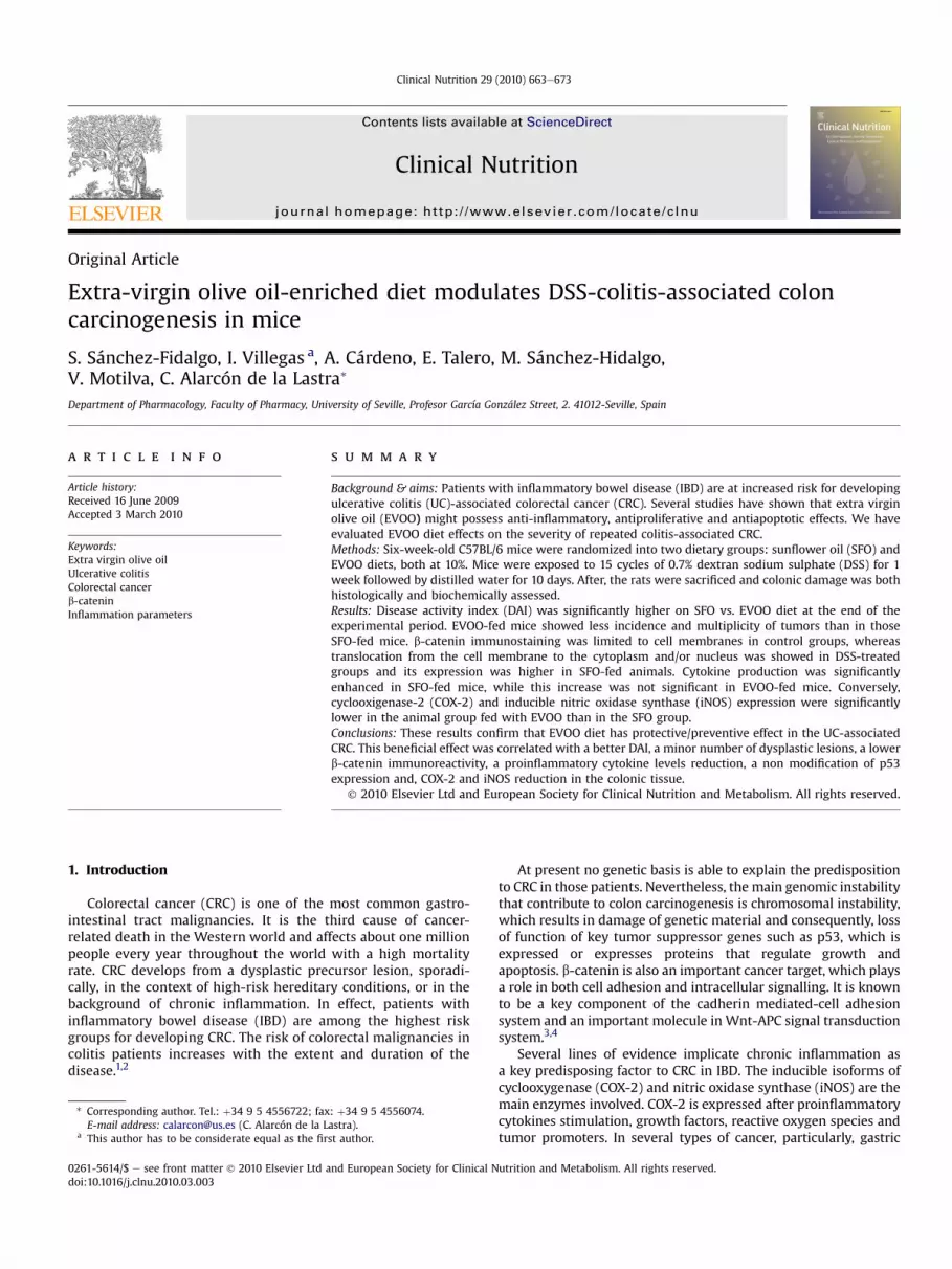
Fig. 1. Overview of experimental protocol in the experimental animal model ofdextran sodium sulphate (DSS)-induced ulcerative colitis model in mice. Chroniculcerative colitis was induced by the repeated administration of 15 cycles of DSS. Eachcycle consisted of 7 days of 0.7% DSS dissolved in the drinking water ad libitum,followed by 10 days of ordinary tap water. Sham group mice were given ordinary tapwater and they were sacrificed at the same time that DSS group.
S. Sánchez-Fidalgo et al. / Clinical Nutrition 29 (2010) 663e673 665
2.3. Assessment of DSS-induced colitis
The clinical activity of colitis was evaluated during experimen-tation in order to determinate the disease activity index (DAI) asdescribed by Gommeaux et al.22 The presence of diarrhea, rectalbleeding, and weight loss were registered at the first, middle andend of DSS treatment as well as the phase of drinking water(for each cycle), and separately graded on a scale (Table 2); theaverage of the three values constituted the DAI.
2.4. Macroscopic and histopathological evaluation
After killing the animals, colons were removed, slightly cleanedin physiological saline to remove fecal residues, weighed andmeasured. The severity of macroscopic damage was evaluated byan independent observer who was blinded to the treatment.Weight/length ratio of the mice colon, as an indirect marker ofinflammation was also determined. Photographs taken from colonsamples were digitised using Kodak D290 Zoom camera (EastmanKodak Co., Rochester, NY, USA). Pieces of colon were collected andfrozen in liquid nitrogen to measure biochemical parameters.
Tissue samples of the region with polypoid or flat elevatedlesion were excised out of every segment, fixed in 4% bufferedformaldehyde, dehydrated by increasing concentrations of ethanol,and embedded in paraffin. The paraffin sections were stained withhaematoxylin and eosin for histological evaluation of colonicdamage. UC-associated colonic neoplasms were analyzed micro-scopically and diagnosed as low-grade or high-grade of dysplasiaand adenocarcinoma, according to established criteria.23 Tumorincidence was calculated as the number of tumor-bearing micedivided by the total number of mice expressed in percentage.Histopathological evaluation was determined by a pathologist whowas unaware of the experimental protocol.
2.5. Immunohistochemical study of b-catenin
b-catenin staining was carried out using a streptavidin-Biotin-Peroxidase method. 5 mm thick tissue sections were driedin an oven at 55 �C for 1 h and then deparaffinized in twochanges of xylene and hydrated from graded alcohols to water. Apressure-cooking antigen retrieval system was used in which the
Table 2Scoring of disease activity index (DAI) in DSS-induced colitis.
Score Bleeding
0 None1 Small spots of blood in stool; dry anal region2 Large spots of blood in stool; blood appears through anal orifice3 Deep red stool; blood spreads largely around the anus
Criteria from work of Gommeaux et al.23
slides were boiled in citrate buffer (pH 6.0) at maximum pres-sure for 2 min, followed by cooling at room temperature for20 min. The endogenous peroxidase activity was inhibited andthen the sections were incubated in normal horse serum(Vectastain� Kit, Vector Laboratories, Inc., Burlingame, CA) for20 min to reduce non-specific staining, and successively incu-bated with monoclonal mouse anti-b-catenin antibody (BDTransduction Laboratories TM) at dilution 1:1500 overnight at4 �C. Subsequently, slides were treated with anti-mouse IgGantibody (Vectastain� Kit, Vector Laboratories, Inc., Burlingame,CA) for 30 min and incubated with the streptavidin-peroxidasecomplex (Vectastain� Kit, Vector Laborartories, Inc., Burlingame,CA) for 30 min, both steps at room temperature. The enzymaticstudy was developed using 3,30ediaminobenzidine (DAB) andthe sections were counterstained with haematoxylin. Negativecontrol sections were treated as the same way omitting theprimary antibody. Intensity and localization of immunoreactivitywas examined on all sections using a microscope (OlympusBX61, Olympus Optical Co., Ltd., Tokyo, Japan). The pathologistevaluated b-catenin staining as percentage of cells in each groupshowing cell membrane or cytoplasmic and/or nucleusexpression.24
2.6. Colonic cytokine levels
TNF-a, IL-6 and IFN-g concentrations in colonic tissue weremeasured by quantitative enzyme immunoassay kits according tothe manufacturer’ s protocol (Diaclone, Besançon, France). Briefly,distal colon samples were weighed and homogenized, after thaw-ing, in 0.3 ml phosphate buffer saline solution (PBS, pH¼ 7.2) 1%bovine serum albumin (BSA) at �C, andwere centrifuged at 12,000 gfor 10 min. The concentrations of cytokines were determined byduplicated. TNF-a, IL-6 and IFN-g values were expressed as pg/mgtissue.
2.7. Isolation of cytoplasmic proteins and Western blot analysis
Frozen colonic tissues were weighed and homogenized in ice-cold buffer (50 mM TriseHCl, pH 7.5, 8 mM MgCl2, 5 mM ethyleneglycol bis (2-aminoethyl ether)-N,N,N,N,-tetraacetic acid, 0.5 mMEDTA, 0.01 mg/ml leupeptin, 0.01 mg/ml pepstatin, 0.01 mg/mlaprotinin, 1 mM phenylmethylsulfonyl fluoride and 250 mM NaCl).Homogenates were centrifuged (12,000 g, 15 min, �C) and thesupernatants were collected and stored at �80 �C. Proteinconcentration of the homogenate was determined followingBradford colorimetric method.25 Aliquots of supernatants con-taining equal amounts of protein (50 mg) were separated on 10%acrylamide gel by sodium dodecyl sulfatepolyacryamide gel elec-trophoresis. In the next step, the proteins were electrophoreticallytransferred onto a nitrocellulose membrane and incubated withspecific primary antibodies: mouse anti-p53 (Ab-1) (Calbiochem,Darmstadt, Germany) at a dilution of 1:20, rabbit anti-COX-2 andrabbit anti-iNOS (Cayman Chemical, Michigan, USA) at a dilution of1:3000 and rabbit anti-PGES-1 (Cayman Chemical, Michigan, USA)at a dilution of 1:500. Each filter was washed three times for 15 min
Weight loss (% of initial wt) Stool consistency
<1 Normal stools1e4.99 Soft pellets not adhering to the anus5e10 Very soft pellets adhering to the anus>10 Liquid stool on long streams; wet anus
S. Sánchez-Fidalgo et al. / Clinical Nutrition 29 (2010) 663e673666
and incubated with the secondary horseradish peroxidase linkedanti-rabbit for COX-2, iNOS and PGES-1 (Pierce Chemical Company,Rockford, IL, USA) or anti-mouse immunoglobulin G for p53 anti-bodies (Santa Cruz Biotechnology, CA). To prove equal loading, theblots were analyzed for b-actin expression using an anti-b-actinantibody (SigmaeAldrich, MO, USA). Immunodetection was per-formed using enhanced chemiluminescence light-detecting kit(SuperSignal1 West Pico Chemiluminescent Substrate, Pierce, IL,USA). Densitometric data were studied following normalization tothe control (house-keeping gene). The signals were analyzed andquantified by a Scientific Imaging Systems (KODAK 1D, ImageAnalysis Software).
2.8. Statistical analysis
All values in the figures and text are expressed as arithmeticmeans� standard error (S.E.M.). Data were evaluated with GraphPad Prism� Version 2.01 software. Comparison were done usingone-way analysis of variance (ANOVA) followed by TukeyeKrameror Dunnett’s test when appropriate. P values of <0.05 wereconsidered statistically significant. In the experiment involvinghistology, immunohistochemistry, or Western blot, the figuresshown are representative of at least six experiments performed ondifferent days.
3. Results
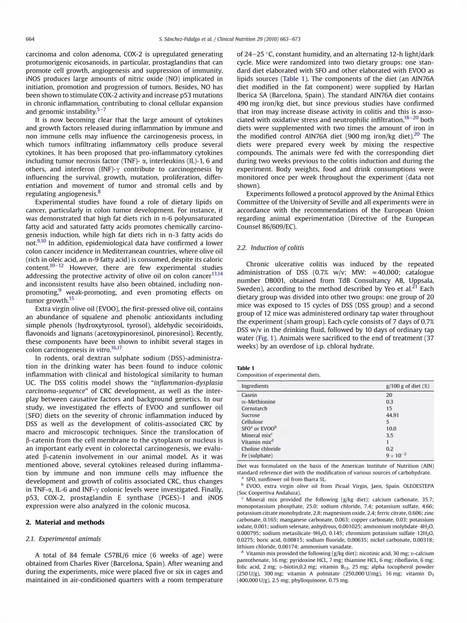
DSSadministration indrinkingwater to theanimals during the15cycles reproduced a chronic colitis in the twodietary groups assayed,SFOandEVOO,withevidentclinic signsofdiarrheaandhaemorrhagerectal. The evolution of body weight and consumption of food anddrink during this period did not show significant differences (datanot show). On the contrary, a significant increase onweight/length ofthemice colonwas observed in the DSS-treated groups and fedwithSFO vs. sham group (P< 0.001), while this difference was notstatistically significant in EVOO-fed animals (Fig. 2).
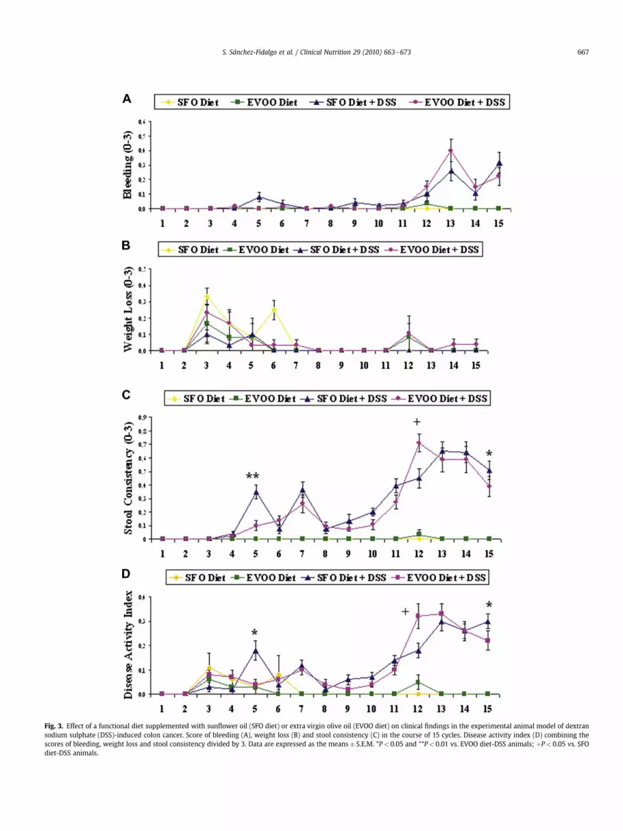
Bleeding examinations were positive and accompanied by loosestools (P< 0.01) from cycle number 5 in SFO-fed animals group. Onthe contrary, visible fecal blood and diarrhea (P< 0.05 vs. SFO diet)were observed from cycle 12 in DSS-EVOO animals. However, DAIshowed fluctuating data from one week to the other between thetwo groups in the course of 15 cycles of DSS treatment, although itwas significantly higher (P< 0.05) on SFO group vs. EVOO diet atthe end of the experimental period (Fig. 3).
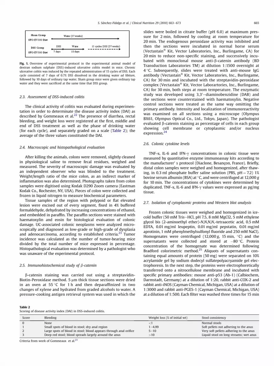
The number, size, and location of detectable tumors weremacroscopically examined. None of the animal controls, SFO orEVOO diets, which were not subject to the cycles of DSS, showedinflammation and/or injury in the colon. A total of 6 out of 20 (30%)
Fig. 2. Effect of a functional diet supplemented with sunflower oil (SFO diet) or extravirgin olive oil (EVOO diet) on weight/length of the colon in the experimental animalmodel of dextran sodium sulphate (DSS)-induced colon cancer. Data are expressed asthe means� S.E.M. ***P< 0.001 vs. Sham group.
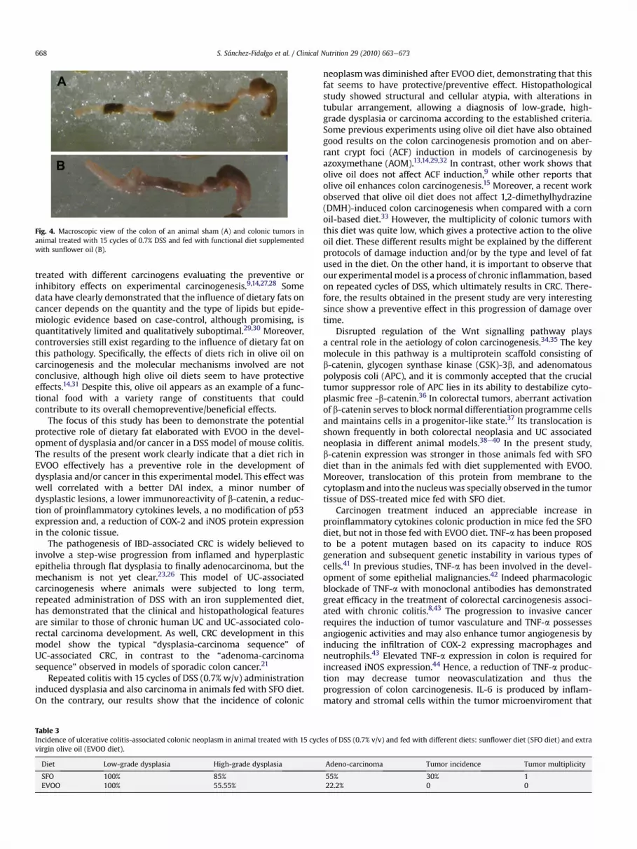
mice that received 15 cycles of DSS and were fed with SFO diet,developed nodular, polypoid or caterpillar-like tumors26 (Fig. 4B).These lesions ranged from 2 to 6 mm in size and appeared in thedistal/rectal portion of the large intestine mainly. In contrast, noneof the animals fed with EVOO diet had any macroscopic lesions.
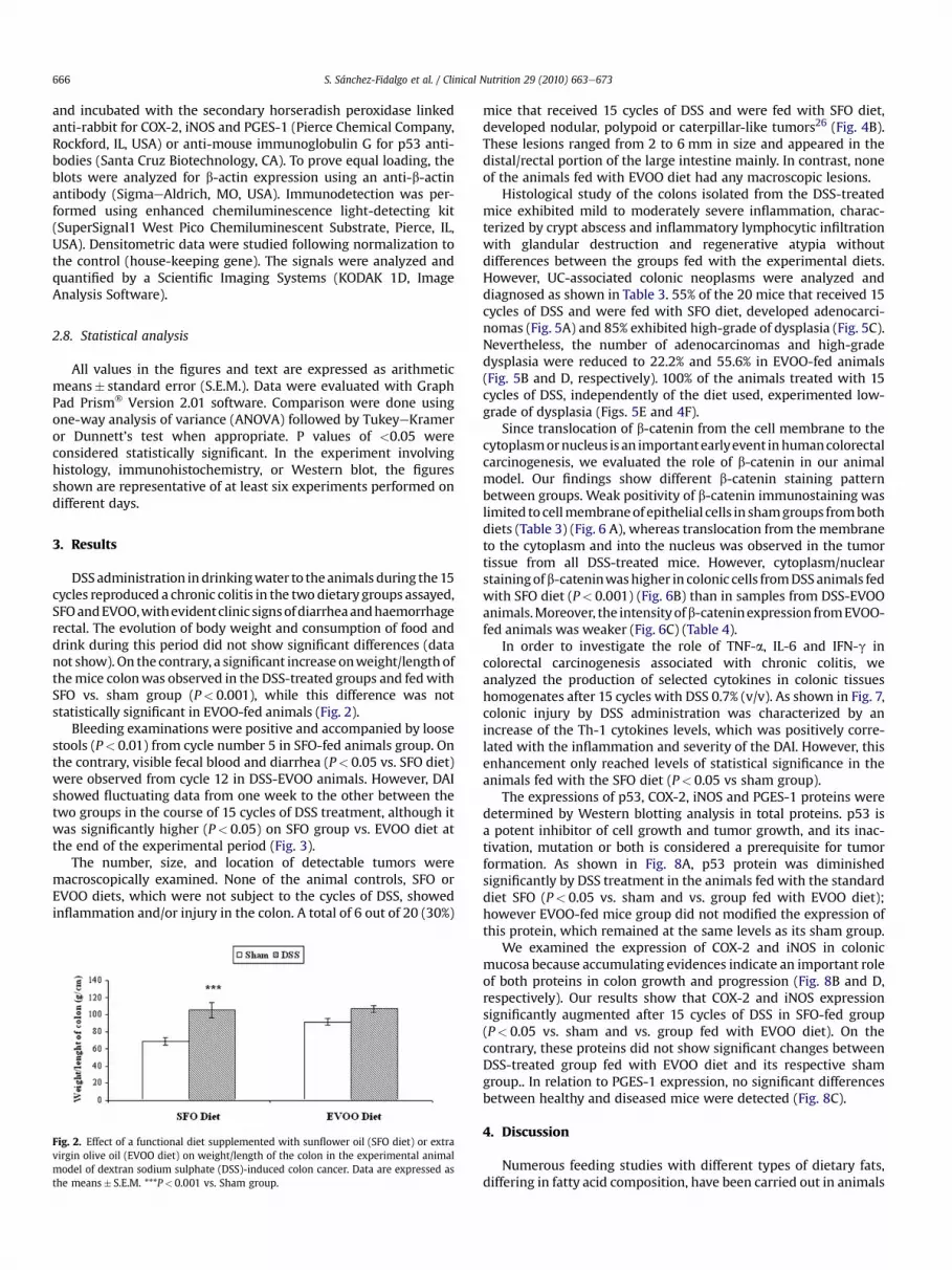
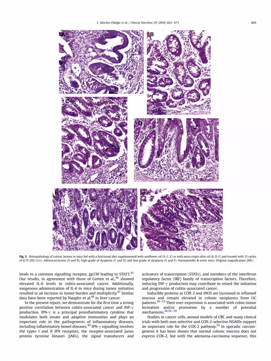
Histological study of the colons isolated from the DSS-treatedmice exhibited mild to moderately severe inflammation, charac-terized by crypt abscess and inflammatory lymphocytic infiltrationwith glandular destruction and regenerative atypia withoutdifferences between the groups fed with the experimental diets.However, UC-associated colonic neoplasms were analyzed anddiagnosed as shown in Table 3. 55% of the 20 mice that received 15cycles of DSS and were fed with SFO diet, developed adenocarci-nomas (Fig. 5A) and 85% exhibited high-grade of dysplasia (Fig. 5C).Nevertheless, the number of adenocarcinomas and high-gradedysplasia were reduced to 22.2% and 55.6% in EVOO-fed animals(Fig. 5B and D, respectively). 100% of the animals treated with 15cycles of DSS, independently of the diet used, experimented low-grade of dysplasia (Figs. 5E and 4F).
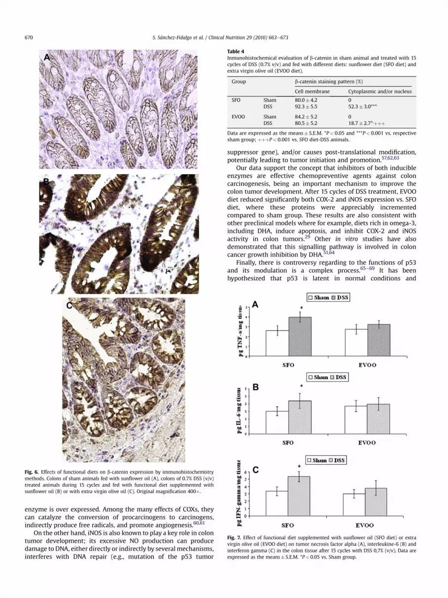
Since translocation of b-catenin from the cell membrane to thecytoplasmornucleus is an importantearlyevent inhumancolorectalcarcinogenesis, we evaluated the role of b-catenin in our animalmodel. Our findings show different b-catenin staining patternbetween groups. Weak positivity of b-catenin immunostaining waslimited to cellmembraneof epithelial cells in shamgroups frombothdiets (Table 3) (Fig. 6 A), whereas translocation from the membraneto the cytoplasm and into the nucleus was observed in the tumortissue from all DSS-treated mice. However, cytoplasm/nuclearstainingofb-cateninwashigher in colonic cells fromDSSanimals fedwith SFO diet (P< 0.001) (Fig. 6B) than in samples from DSS-EVOOanimals.Moreover, the intensityofb-cateninexpression fromEVOO-fed animals was weaker (Fig. 6C) (Table 4).
In order to investigate the role of TNF-a, IL-6 and IFN-g incolorectal carcinogenesis associated with chronic colitis, weanalyzed the production of selected cytokines in colonic tissueshomogenates after 15 cycles with DSS 0.7% (v/v). As shown in Fig. 7,colonic injury by DSS administration was characterized by anincrease of the Th-1 cytokines levels, which was positively corre-lated with the inflammation and severity of the DAI. However, thisenhancement only reached levels of statistical significance in theanimals fed with the SFO diet (P< 0.05 vs sham group).
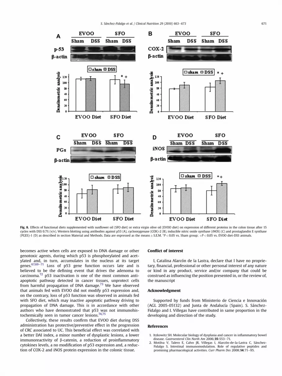
The expressions of p53, COX-2, iNOS and PGES-1 proteins weredetermined by Western blotting analysis in total proteins. p53 isa potent inhibitor of cell growth and tumor growth, and its inac-tivation, mutation or both is considered a prerequisite for tumorformation. As shown in Fig. 8A, p53 protein was diminishedsignificantly by DSS treatment in the animals fed with the standarddiet SFO (P< 0.05 vs. sham and vs. group fed with EVOO diet);however EVOO-fed mice group did not modified the expression ofthis protein, which remained at the same levels as its sham group.
We examined the expression of COX-2 and iNOS in colonicmucosa because accumulating evidences indicate an important roleof both proteins in colon growth and progression (Fig. 8B and D,respectively). Our results show that COX-2 and iNOS expressionsignificantly augmented after 15 cycles of DSS in SFO-fed group(P< 0.05 vs. sham and vs. group fed with EVOO diet). On thecontrary, these proteins did not show significant changes betweenDSS-treated group fed with EVOO diet and its respective shamgroup.. In relation to PGES-1 expression, no significant differencesbetween healthy and diseased mice were detected (Fig. 8C).
4. Discussion
Numerous feeding studies with different types of dietary fats,differing in fatty acid composition, have been carried out in animals
Fig. 3. Effect of a functional diet supplemented with sunflower oil (SFO diet) or extra virgin olive oil (EVOO diet) on clinical findings in the experimental animal model of dextransodium sulphate (DSS)-induced colon cancer. Score of bleeding (A), weight loss (B) and stool consistency (C) in the course of 15 cycles. Disease activity index (D) combining thescores of bleeding, weight loss and stool consistency divided by 3. Data are expressed as the means� S.E.M. *P< 0.05 and **P< 0.01 vs. EVOO diet-DSS animals; þP< 0.05 vs. SFOdiet-DSS animals.
S. Sánchez-Fidalgo et al. / Clinical Nutrition 29 (2010) 663e673 667
Fig. 4. Macroscopic view of the colon of an animal sham (A) and colonic tumors inanimal treated with 15 cycles of 0.7% DSS and fed with functional diet supplementedwith sunflower oil (B).
S. Sánchez-Fidalgo et al. / Clinical Nutrition 29 (2010) 663e673668
treated with different carcinogens evaluating the preventive orinhibitory effects on experimental carcinogenesis.9,14,27,28 Somedata have clearly demonstrated that the influence of dietary fats oncancer depends on the quantity and the type of lipids but epide-miologic evidence based on case-control, although promising, isquantitatively limited and qualitatively suboptimal.29,30 Moreover,controversies still exist regarding to the influence of dietary fat onthis pathology. Specifically, the effects of diets rich in olive oil oncarcinogenesis and the molecular mechanisms involved are notconclusive, although high olive oil diets seem to have protectiveeffects.14,31 Despite this, olive oil appears as an example of a func-tional food with a variety range of constituents that couldcontribute to its overall chemopreventive/beneficial effects.
The focus of this study has been to demonstrate the potentialprotective role of dietary fat elaborated with EVOO in the devel-opment of dysplasia and/or cancer in a DSS model of mouse colitis.The results of the present work clearly indicate that a diet rich inEVOO effectively has a preventive role in the development ofdysplasia and/or cancer in this experimental model. This effect waswell correlated with a better DAI index, a minor number ofdysplastic lesions, a lower immunoreactivity of b-catenin, a reduc-tion of proinflammatory cytokines levels, a no modification of p53expression and, a reduction of COX-2 and iNOS protein expressionin the colonic tissue.
The pathogenesis of IBD-associated CRC is widely believed toinvolve a step-wise progression from inflamed and hyperplasticepithelia through flat dysplasia to finally adenocarcinoma, but themechanism is not yet clear.23,26 This model of UC-associatedcarcinogenesis where animals were subjected to long term,repeated administration of DSS with an iron supplemented diet,has demonstrated that the clinical and histopathological featuresare similar to those of chronic human UC and UC-associated colo-rectal carcinoma development. As well, CRC development in thismodel show the typical “dysplasia-carcinoma sequence” ofUC-associated CRC, in contrast to the “adenoma-carcinomasequence” observed in models of sporadic colon cancer.21
Repeated colitis with 15 cycles of DSS (0.7% w/v) administrationinduced dysplasia and also carcinoma in animals fed with SFO diet.On the contrary, our results show that the incidence of colonic
Table 3Incidence of ulcerative colitis-associated colonic neoplasm in animal treated with 15 cyclvirgin olive oil (EVOO diet).
Diet Low-grade dysplasia High-grade dysplasia
SFO 100% 85%EVOO 100% 55.55%
neoplasmwas diminished after EVOO diet, demonstrating that thisfat seems to have protective/preventive effect. Histopathologicalstudy showed structural and cellular atypia, with alterations intubular arrangement, allowing a diagnosis of low-grade, high-grade dysplasia or carcinoma according to the established criteria.Some previous experiments using olive oil diet have also obtainedgood results on the colon carcinogenesis promotion and on aber-rant crypt foci (ACF) induction in models of carcinogenesis byazoxymethane (AOM).13,14,29,32 In contrast, other work shows thatolive oil does not affect ACF induction,9 while other reports thatolive oil enhances colon carcinogenesis.15 Moreover, a recent workobserved that olive oil diet does not affect 1,2-dimethylhydrazine(DMH)-induced colon carcinogenesis when compared with a cornoil-based diet.33 However, the multiplicity of colonic tumors withthis diet was quite low, which gives a protective action to the oliveoil diet. These different results might be explained by the differentprotocols of damage induction and/or by the type and level of fatused in the diet. On the other hand, it is important to observe thatour experimental model is a process of chronic inflammation, basedon repeated cycles of DSS, which ultimately results in CRC. There-fore, the results obtained in the present study are very interestingsince show a preventive effect in this progression of damage overtime.
Disrupted regulation of the Wnt signalling pathway playsa central role in the aetiology of colon carcinogenesis.34,35 The keymolecule in this pathway is a multiprotein scaffold consisting ofb-catenin, glycogen synthase kinase (GSK)-3b, and adenomatouspolyposis coli (APC), and it is commonly accepted that the crucialtumor suppressor role of APC lies in its ability to destabilize cyto-plasmic free -b-catenin.36 In colorectal tumors, aberrant activationof b-catenin serves to block normal differentiation programme cellsand maintains cells in a progenitor-like state.37 Its translocation isshown frequently in both colorectal neoplasia and UC associatedneoplasia in different animal models.38e40 In the present study,b-catenin expression was stronger in those animals fed with SFOdiet than in the animals fed with diet supplemented with EVOO.Moreover, translocation of this protein from membrane to thecytoplasm and into the nucleus was specially observed in the tumortissue of DSS-treated mice fed with SFO diet.
Carcinogen treatment induced an appreciable increase inproinflammatory cytokines colonic production in mice fed the SFOdiet, but not in those fed with EVOO diet. TNF-a has been proposedto be a potent mutagen based on its capacity to induce ROSgeneration and subsequent genetic instability in various types ofcells.41 In previous studies, TNF-a has been involved in the devel-opment of some epithelial malignancies.42 Indeed pharmacologicblockade of TNF-a with monoclonal antibodies has demonstratedgreat efficacy in the treatment of colorectal carcinogenesis associ-ated with chronic colitis.8,43 The progression to invasive cancerrequires the induction of tumor vasculature and TNF-a possessesangiogenic activities and may also enhance tumor angiogenesis byinducing the infiltration of COX-2 expressing macrophages andneutrophils.43 Elevated TNF-a expression in colon is required forincreased iNOS expression.44 Hence, a reduction of TNF-a produc-tion may decrease tumor neovasculatization and thus theprogression of colon carcinogenesis. IL-6 is produced by inflam-matory and stromal cells within the tumor microenviroment that
es of DSS (0.7% v/v) and fed with different diets: sunflower diet (SFO diet) and extra
Fig. 5. Histopathology of colonic lesions in mice fed with a functional diet supplemented with sunflower oil (A, C, E) or with extra virgin olive oil (B, D, F) and treated with 15 cyclesof 0.7% DSS (v/v). Adenocarcinoma (A and B), high-grade of dysplasia (C and D) and low-grade of dysplasia (E and F). Haematoxilin & eosin stain. Original magnification 200�.
S. Sánchez-Fidalgo et al. / Clinical Nutrition 29 (2010) 663e673 669
binds to a common signalling receptor, gp130 leading to STAT3.45
Our results, in agreement with those of Greten et al.,46 showedelevated IL-6 levels in colitis-associated cancer. Additionally,exogenous administration of IL-6 to mice during tumor initiationresulted in an increase in tumor burden and multiplicity.47 Similardata have been reported by Naugler et al.48 in liver cancer.
In the present report, we demonstrate for the first time a strongpositive correlation between colitis-associated cancer and INF-gproduction. IFN-g is a principal proinflammatory cytokine thatmodulates both innate and adaptive immunities and plays animportant role in the pathogenesis of inflammatory diseases,including inflammatory bowel diseases.49 IFN-g signalling involvesthe types I and II IFN receptors, the receptor-associated Janusprotein tyrosine kinases (JAKs), the signal transducers and
activators of transcription (STATs), and members of the interferonregulatory factor (IRF) family of transcription factors. Therefore,reducing INF-g production may contribute to retard the initiationand progression of colitis-associated cancer.
Inducible proteins as COX-2 and iNOS are increased in inflamedmucosa and remain elevated in colonic neoplasms from UCpatients.50e53 Their over expression is associated with colon tumorformation and/or promotion by a number of potentialmechanisms.28,54e59
Studies in cancer cells, animal models of CRC and many clinicaltrials with both non-selective and COX-2-selective NSAIDs supportan important role for the COX-2 pathway.54 In sporadic carcino-genesis it has been shown that normal colonic mucosa does notexpress COX-2, but with the adenoma-carcinoma sequence, this
Fig. 6. Effects of functional diets on b-catenin expression by immunohistochemistrymethods. Colons of sham animals fed with sunflower oil (A), colons of 0.7% DSS (v/v)treated animals during 15 cycles and fed with functional diet supplemented withsunflower oil (B) or with extra virgin olive oil (C). Original magnification 400�.
Table 4Inmunohistochemical evaluation of b-catenin in sham animal and treated with 15cycles of DSS (0.7% v/v) and fed with different diets: sunflower diet (SFO diet) andextra virgin olive oil (EVOO diet).
Group b-catenin staining pattern (%)
Cell membrane Cytoplasmic and/or nucleus
SFO Sham 80.0� 4.2 0DSS 92.3� 5.5 52.3� 3.0***
EVOO Sham 84.2� 5.2 0DSS 80.5� 5.2 18.7� 2.7*,þþþ
Data are expressed as the means� S.E.M. *P< 0.05 and ***P< 0.001 vs. respectivesham group; þþþP< 0.001 vs. SFO diet-DSS animals.
Fig. 7. Effect of functional diet supplemented with sunflower oil (SFO diet) or extravirgin olive oil (EVOO diet) on tumor necrosis factor alpha (A), interleukine-6 (B) andinterferon gamma (C) in the colon tissue after 15 cycles with DSS 0,7% (v/v). Data areexpressed as the means� S.E.M. *P< 0.05 vs. Sham group.
S. Sánchez-Fidalgo et al. / Clinical Nutrition 29 (2010) 663e673670
enzyme is over expressed. Among the many effects of COXs, theycan catalyze the conversion of procarcinogens to carcinogens,indirectly produce free radicals, and promote angiogenesis.60,61
On the other hand, iNOS is also known to play a key role in colontumor development; its excessive NO production can producedamage to DNA, either directly or indirectly by several mechanisms,interferes with DNA repair (e.g., mutation of the p53 tumor
suppressor gene), and/or causes post-translational modification,potentially leading to tumor initiation and promotion.57,62,63
Our data support the concept that inhibitors of both inducibleenzymes are effective chemopreventive agents against coloncarcinogenesis, being an important mechanism to improve thecolon tumor development. After 15 cycles of DSS treatment, EVOOdiet reduced significantly both COX-2 and iNOS expression vs. SFOdiet, where these proteins were appreciably incrementedcompared to sham group. These results are also consistent withother preclinical models where for example, diets rich in omega-3,including DHA, induce apoptosis, and inhibit COX-2 and iNOSactivity in colon tumors.29 Other in vitro studies have alsodemonstrated that this signalling pathway is involved in coloncancer growth inhibition by DHA.51,64
Finally, there is controversy regarding to the functions of p53and its modulation is a complex process.65e69 It has beenhypothesized that p53 is latent in normal conditions and
Fig. 8. Effects of functional diets supplemented with sunflower oil (SFO diet) or extra virgin olive oil (EVOO diet) on expression of different proteins in the colon tissue after 15cycles with DSS 0.7% (v/v). Western blotting using antibodies against p53 (A), cyclooxygenase (COX)-2 (B), inducible nitric oxide synthase (iNOS) (C) and prostaglandin E synthase(PGES)-1 (D) as described in section Material and Methods. Data are expressed as the means� S.E.M. *P< 0.05 vs. Sham group; þP< 0.05 vs. EVOO diet-DSS animals.
S. Sánchez-Fidalgo et al. / Clinical Nutrition 29 (2010) 663e673 671
becomes active when cells are exposed to DNA damage or othergenotoxic agents, during which p53 is phosphorylated and acet-ylated and, in turn, accumulates in the nucleus at its targetgenes.67,69e71 Loss of p53 gene function occurs late and isbelieved to be the defining event that drives the adenoma tocarcinoma.72 p53 inactivation is one of the most common anti-apoptotic pathway detected in cancer tissues, unprotect cellsfrom harmful propagation of DNA damage.73 We have observedthat animals fed with EVOO did not modify p53 expression and,on the contrary, loss of p53 function was observed in animals fedwith SFO diet, which may inactive apoptotic pathway driving topropagation of DNA damage. This is in accordance with otherauthors who have demonstrated that p53 was not immunohis-tochemically seen in tumor cancer lesions.74,75
Collectively, these results confirm that EVOO diet during DSSadministration has protective/preventive effect in the progressionof CRC associated to UC. This beneficial effect was correlated witha better DAI index, a minor number of dysplastic lesions, a lowerimmunoreactivity of b-catenin, a reduction of proinflammatorycytokines levels, a no modification of p53 expression and, a reduc-tion of COX-2 and iNOS protein expression in the colonic tissue.
Conflict of interest
I, Catalina Alarcón de la Lastra, declare that I have no proprie-tary, financial, professional or other personal interest of any natureor kind in any product, service and/or company that could beconstrued as influencing the position presented in, or the review of,the manuscript
Acknowledgment
Supported by funds from Ministerio de Ciencia e Innovación(AGL 2005-05132) and Junta de Andalucía (Spain). S. Sánchez-Fidalgo and I. Villegas have contributed in same proportion in thedeveloping and direction of the study.
References
1. Itzkowitz SH. Molecular biology of dysplasia and cancer in inflammatory boweldisease. Gastroenterol Clin North Am 2006;35:553e71.
2. Motilva V, Talero E, Calvo JR, Villegas I, Alarcón-de-la-Lastra C, Sánchez-Fidalgo S. Intestinal immunomodulation. Role of regulative peptides andpromising pharmacological activities. Curr Pharm Des 2008;14:71e95.
S. Sánchez-Fidalgo et al. / Clinical Nutrition 29 (2010) 663e673672
3. Gavert N, Ben-Ze’ev A. Beta-Catenin signaling in biological control and cancer.J Cell Biochem 2007;102:820e8.
4. Tanaka T, Kohno H, Suzuki R, Yamada Y, Sugie S, Mori H. A novel inflammation-related mouse colon carcinogenesis model induced by azoxymethane anddextran sodium sulfate. Cancer Sci 2003;94:965e73.
5. Müller-Decker K, Fürstenberger G. The cyclooxygenase-2-mediated prosta-glandin signaling is causally related to epithelial carcinogenesis. Mol Carcinog2007;46:705e10.
6. Kawanishi S, Hiraku Y, Pinlaor S, Ma N. Oxidative and nitrative DNA damage inanimals and patients with inflammatory diseases in relation to inflammation-related carcinogenesis. Biol Chem 2006;387:365e72.
7. Mutoh M, Takahashi M, Wakabayashi K. Roles of prostanoids in coloncarcinogenesis and their potential targeting for cancer chemoprevention.Curr Pharm Des 2006;12:2375e82.
8. Burstein E, Fearon ER. Colitis and cancer: a tale of inflammatory cells and theircytokines. J Clin Invest 2008;118:464e7.
9. Davidson LA, Nguyen DV, Hokanson RM, Callaway ES, Isett RB, Turner ND, et al.Chemopreventive n-3 polyunsaturated fatty acids reprogram genetic signaturesduring colon cancer initiation and progression in the rat. Cancer Res2004;64:6797e804.
10. Owen RW, Haubner R, Wurtele G, Hull E, Spiegelhalder B, Bartsch H. Olives andolive oil in cancer prevention. Eur J Cancer Prev 2004;13:319e26.
11. Cottet V, Bonithon-Kopp C, Kronborg O, Santos L, Andreatta R, Boutron-Ruault MC, et al, European Cancer Prevention Organisation Study Group.Dietary patterns and the risk of colorectal adenoma recurrence in a Europeanintervention trial. Eur J Cancer Prev 2005;14:21e9.
12. Alarcón de la Lastra C, Barranco MD, Motilva V, Herrerias JM. Mediterraneandiet and health: biological importance of olive oil. Curr Pharm Des2001;7:933e50.
13. Fujise T, Iwakiri R, Kakimoto T, Shiraishi R, Sakata Y, Wu B, et al. Long-termfeeding of various fat diets modulates azoxymethane-induced colon carcino-genesis through Wnt/beta-catenin signaling in rats. Am J Physiol GastrointestLiver Physiol 2007;292:G1150eG1156.
14. Bartoli R, Fernandez-Banares F, Navarro E, Castella E, Mane J, Alvarez M, et al.Effect of olive oil on early and late events of colon carcinogenesis in rats:modulation of arachidonic acid metabolism and local prostaglandin (E2)synthesis. Gut 2000;46:191e9.
15. Onogi N, Okuno M, Komaki C, Moriwaki H, Kawamori T, Tanaka T, et al.Suppressing effect of perilla oil on azoxymethane-induced foci of colonicaberrant crypts in rats. Carcinogenesis 1996;17:1291e6.
16. Hashim YZ, Rowland IR, McGlynn H, Servili M, Selvaggini R, Taticchi A, et al.Inhibitory effects of olive oil phenolics on invasion in human colon adenocar-cinoma cells in vitro. Int J Cancer 2008;122:495e500.
17. Gill CI, Boyd A, McDermott E, McCann M, Servili M, Selvaggini R, et al. Potentialanti-cancer effects of virgin olive oil phenols on colorectal carcinogenesismodels in vitro. Int J Cancer 2005;117:1e7.
18. Carrier JC, Aghdassi E, Jeejeebhoy K, Allard JP. Exacerbation of dextran sulfatesodium-induced colitis by dietary iron supplementation: role of NF-kappaB.Int J Colorectal Dis 2006;21:81e387.
20. Seril DN, Liao J, YangCS, YangGY. Systemic iron supplementation replenishes ironstores without enhancing colon carcinogenesis in murine models of ulcerativecolitis: comparison with iron-enriched diet. Dig Dis Sci 2005;50:696e707.
21. Yeo M, Kim DK, Park HJ, Oh TY, Kim JH, Cho SW, et al. Loss of transgelin inrepeated bouts of ulcerative colitis-induced colon carcinogenesis. Proteomics2006;6:1158e65.
22. Gommeaux J, Cano C, Garcia S, Gironella M, Pietri S, Culcasi M, et al. Colitis andcolitis-associated cancer are exacerbated in mice deficient for tumor protein53-induced nuclear protein 1. Mol Cell Biol 2007;27:2215e28.
24. Osawa E, Nakajima A, Fujisawa T, et al. Predominant T helper type 2-inflam-matory responses promote murine colon cancers. Int J Cancer2006;118:2232e6.
25. Bradford MM. A rapid and sensitive method for the quantitation of microgramquantities of protein utilizing the principle of proteinedye binding. Anal Bio-chem 1976;72:248e54.
26. Kohno H, Suzuki R, Sugie S, Tanaka T. Suppression of colitis-related mousecolon carcinogenesis by a COX-2 inhibitor and PPAR ligands. BMC Cancer2005;5:46.
27. Woutersen RA, Appel MJ, Garderen-Hoetmer A, Wijnands M. Dietary fat andcarcinogenesis. Mutat Res 1999;443:111e27.
29. Rao CV, Hirose Y, Indranie C, Reddy BS. Moduration of experimental colontumorigenesis by types and amounts of dietary fatty acids. Cancer Res2001;61:1927e33.
30. Lipworth L, Martinez ME, Angell J, Hsieh CC, Trichopoulos D. Olive oil andhuman cancer: an assessment of the evidence. Prev Med 1997;26:181e90.
31. Escrich E, Moral R, Grau L, Costa I, Solanas M. Molecular mechanisms of theeffects of olive oil and other dietary lipids on cancer. Mol Nutr Food Res2007;51:1279e92.
32. Schwartz B, Birk Y, Raz A, Madar Z. Nutritional-pharmacological combinations:a novel approach to reducing colon cancer incidence. Eur J Nutr2004;43:221e9.
33. Femia A, Dolara P, Servili M, Esposto S, Taticchi A, Urbani S, et al. No effects ofolive oils with different phenolic content compared to corn oil on 1,2-dimethylhydrazine-induced colon carcinogenesis in rats. Eur J Nutr 2008;47:329e334.
34. Liu W, Dong X, Mai M, Seelan RS, Taniguchi K, Krishnandath KK, Halling KC,Cunningham JM, Boardman LA, Qian C, Christensen E, Schmidt SS, Roche PC,Smith DI, Thibodeau SN. Mutations in Axin2 cause colorectal cancer withdetective mismatch repair by activating beta-c-atenin-Tcf signaling. Nat Genet2000;26:146e7.
35. Morin PJ, Sparks AB, Korinek V, Barker N, Clevers H, Vogelstein B, et al.Activation of beta-catenin-Tcf signaling in colon cancer by mutation in betacatenin or APC. Science 1997;275:1787e90.
36. von Kries JP, Winbeck G, Asbrand C, Schwarz-Romond T, Sochnikowa N,Dell’Oro A, Behrens J, Birchmeier W. Hot spots in betacatenin for interactionswith LEF-1, conduction and APC. Nat Struct Biol 2000;7:800e7.
37. van de Wetering M, Sancho E, Verweij C, de Lau W, Oving I, Hurlstone A, et al.The beta-catenin/TCF-4 complex imposes a crypt progenitor phenotype oncolorectal cancer cells. Cell 2002;111:241e50.
38. Fujii S, Fujimori T, Kawamata H, Takada J, Kitajima K, Omotehara F, Kaihara T,Kusaka T, Ichikawa K, Ohkura Y, Ono Y, Imura J, Yamaoka S, Sakamoto C,Ueda Y, Chiba T. Development of colonic neoplasia in p53 deficient mice withexperimental colitis induced by dextran sulphate sodium. Gut 2004;53:710e6.
39. Furihata T, Kawamata H, Kubota K, Fujimori T. Evaluation of the malignantpotential of aberrant crypt foci by immunohistochemical staining for betaca-tenin in inflammation-induced rat colon carcinogenesis. Int J Mol Med2002;9:353e8.
40. Aust DE, Terdiman JP, Willenbucher RF, Chew K, Ferrell L, Florendo C, Molinaro-Clark A, Baretton GB, Löhrs U, Waldman FM. Altered distribution of beta-cat-enin, and its binding proteins E-cadherin and APC, in ulcerative colitis-relatedcolorectal cancers. Mod Pathol 2001;14:29e39.
41. Babbar N, Casero Jr RA. Tumor necrosis factor-alpha increases reactive oxygenspecies by inducing spermine oxidase in human lung epithelial cells: a poten-tial mechanism for inflammation-induced carcinogenesis. Cancer Res2006;66:11,125e11,130.
42. Scott KA, Moore RJ, Arnott CH, East N, Thompson RG, Scallon BJ, Shealy DJ,Balkwill FR. An anti-tumor necrosis factor-alpha antibody inhibits the devel-opment of experimental skin tumors. Mol Cancer Ther 2003;2:445e51.
43. Popivanova BK, Kitamura K, Wu Y, Kondo T, Kagaya T, Kaneko S, et al. BlockingTNF-alpha in mice reduces colorectal carcinogenesis associated with chroniccolitis. J Clin Invest 2008;118:560e70.
44. Erdman SE, Rao VP, Poutahidis T, Rogers AB, Taylor CL, Jackson EA, Ge Z, Lee CW,Schauer DB, Wogan GN, Tannenbaum SR, Fox JG. Nitric oxide and TNF-alphatrigger colonic inflammation and carcinogenesis in Helicobacter hepaticus-infected, Rag2-deficient mice. Proc Natl Acad Sci USA 2009;106:1027e32.
45. Bromberg J, Wang TC. Inflammation and cancer: IL-6 and STAT3 complete thelink. Cancer Cell 2009;15:79e80.
46. Greten FR, Eckmann L, Greten TF, Park JM, Li ZW, Egan LJ, Kagnoff MF, Karin M.IKKbeta links inflammation and tumorigenesis in a mouse model of colitis-associated cancer. Cell 2004;118:285e96.
47. Grivennikov S, Karin E, Terzic J, Mucida D, Yu GY, Vallabhapurapu S, et al. IL-6and stat3 are required for survival of intestinal epithelial cells and developmentof colitis-associated cancer. Cancer Cell. 2009;15:103e13.
48. Naugler WE, Sakurai T, Kim S, Maeda S, Kim K, Elsharkawy AM, Karin M.Gender disparity in liver cancer due to sex differences in MyD88-dependentIL-6 production. Science 2007;317:121e4.
49. Bouma G, Strober W. The immunological and genetic basis of inflammatorybowel disease. Nat Rev Immunol 2003;3:521e33.
50. Itzkowitz SH, Yio X. Inflammation and cancer IV. Colorectal cancer in inflam-matory bowel disease: the role of inflammation. Am J Physiol Gastrointest LiverPhysiol 2004;287:G7eG17.
51. Narayanan BA, Narayanan NK, Simi B, Reddy BS. Modulation of inducible nitricoxide synthase and related proinflammatory genes by the omega-3 fatty aciddocosahexaenoic acid in human colon cancer cells. Cancer Res 2003;63:972e9.
52. Elder DJ, Baker JA, Banu NA, Moorghen M, Paraskeva C. Human colorectaladenomas demonstrate a size-dependent increase in epithelial cyclo-oxygenase-2 expression. J Pathol 2002;198:428e34.
53. Rao CV, Indranie C, Simi B, Manning PT, Connor JR, Reddy BS. Chemopreventiveproperties of a selective inducible nitric oxide synthase inhibitor in coloncarcinogenesis, administered alone or in combination with celecoxib, a selec-tive cyclooxygenase-2 inhibitor. Cancer Res 2002;62:165e70.
54. Greenhough A, Smartt HJ, Moore AE, Roberts HR, Williams AC, Paraskeva C,Kaidi A. The COX-2/PGE2 pathway: key roles in the hallmarks of cancerand adaptation to the tumor microenvironment. Carcinogenesis 2009;30:377e386.
55. Samanta S, Swamy V, Suresh D, Rajkumar M, Rana B, Rana A, et al. Protectiveeffects of vanadium against DMH-induced genotoxicity and carcinogenesis inrat colon: removal of O6-methylguanine DNA adducts, p53 expression,inducible nitric oxide synthase downregulation and apoptotic induction. MutatRes 2008;650:123e31.
56. Dempke W, Rie C, Grothey A, Schmoll HJ. Cyclooxygenase-2: a novel target forcancer chemotherapy? J Cancer Res Clin Oncol 2001;127:411e7.
S. Sánchez-Fidalgo et al. / Clinical Nutrition 29 (2010) 663e673 673
57. Lala PK, Chakraborty C. Role of nitric oxide in carcinogenesis and tumourprogression. Lancet Oncol 2001;2:149e56.
58. Williams CS, Mann M, DuBois RN. The role of cyclooxygenases in inflammation,cancer, and development. Oncogene 1999;18:7908e16.
59. Battu S, Rigaud M, Beneytout JL. Resistance to apoptosis and cyclooxygenase-2expression in a human adenocarcinoma cell line HT29 CL.19A. Anticancer Res1998;18:3579e83.
60. Steele VE, Hawk ET, Viner JL, Lubet RA. Mechanisms and applications of non-steroidal anti-inflammatory drugs in the chemoprevention of cancer. Mutat Res2003;523e524:137e44.
61. Dannenberg AJ, Zakim D. Chemoprevention of colorectal cancer through inhi-bition of cyclooxygenase-2. Semin Oncol 1999;26:499e504.
62. Felley-Bosco E. Role of nitric oxide in genotoxicity: implication for carcino-genesis. Cancer Metastasis Rev 1998;17:25e37.
63. Jenkins DC, Charles IG, Thomsen LL, Moss DW, Holmes LS, Baylis SA, et al. Rolesof nitric oxide in tumor growth. Proc Natl Acad Sci USA 1995;92:4392e6.
64. Narayanan BA, Narayanan NK, Reddy BS. Docosahexaenoic acid (DHA) regu-lated genes and transcription factors inducing apoptosis in human colon cancercells. Int J Oncol 2001;19:1255e62.
65. Barlev NA, Liu L, Chehab NH, Mansfield K, Harris KG, Halazonetis TD, et al.Acetylation of p53 activates transcription through recruitment of coactivators/histone acetyltransferases. Mol Cell 2001;8:243e54.
66. Espinosa JM, Emerson BM. Transcriptional regulation by p53 through intrinsicDNA/chromatin binding and site-directed cofactor recruitment. Mol Cell2001;8:57e69.
67. Liu L, Scolnick D, Trievel RC, Zhang HB, Marmorstein R, Halazonetis TD, et al.p53 sites acetylated in vitro by PCAF and p300 are acetylated in vivo inresponse to DNA damage. Mol Cell Biol 1999;19:1202e9.
68. Sakaguchi K, Herrera JE, Saito S, Miki T, Bustin M, Vassilev A, Anderson CW,Appella E. DNA damage activates p53 through a phosphorylation-acetylationcascade. Genes Dev 1998;12:2831e41.
69. Gu W, Roeder R. Activation of p53 sequence-specific DNA binding by acetyla-tion of the p53 C-terminal domain. Cell 1997;90:595e606.
70. Takenaka I, Morin FF, Seizinger BR, Kley N. Regulation of the sequence-specificDNA binding function of p53 by protein kinase C and protein phosphatases.J Biol Chem 1995;270:5405e11.
71. Hupp TR, Lane DP. Allosteric activation of latent p53 tetramers. Curr Biol1994;4:865e75.
72. Xie J, Itzkowitz SH. Cancer in inflammatory bowel disease.World J Gastroenterol2008;14:378e89.
73. Fridman JS, Lowe SW. Control of apoptosis by p53. Oncogene 2003;22:9030e9040.
74. Wang JG, Wang DF, Lv BJ, Si JM. A novel mouse model for colitis-associatedcolon carcinogenesis induced by 1,2 dimethylhydrazine and dextran sulfatesodium. World J Gastroenterol 2004;10:2958e62.
75. Cooper HS, Murthy S, Kido K, Yoshitake H, Flanigan A. Dysplasia andcancer in the dextran sulfate sodium mouse colitis model. Relevance tocolitis-associated neoplasia in the human: a study of histopathology,B-catenin and p53 expression and the role of inflammation. Carcinogenesis2000;21:757e68.